Background Perforation of an acute ulcer is a serious and life-threatening condition. Treatment for this sudden abdominal event is surgical in the majority of cases.
Image galleries
Literature & Galleries
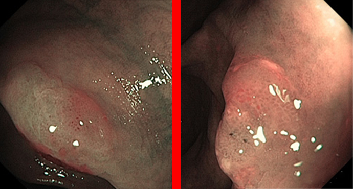
Endoscopic treatment for peptic ulcer perforation

Rapunzel syndrome
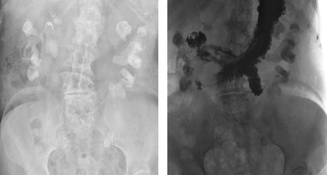
A 31-year-old female patient presented to the University Hospital’s emergency department with upper abdominal pain that had persisted for 2 weeks. She also reported a
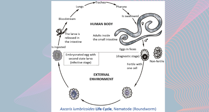
Ascaris Lumbricoides worm encountered in the stomach
Ascaris infestation show quite a wide array of clinical manifestations as it maybe asymptomatic showing only long term manifestations of growth retardation and malnutrition ,
Transcutaneous SpyGlass-guided rendezvous
Achieving sustained biliary drainage in patients with cholestasis or cholangitis often represents a turning-point in the treatment of malignant and infectious diseases with hepatobiliary involvement.

Endoscopic therapy in sigmoid volvulus — case report and glance at the current ASGE guideline
Clinical case: A 78-year-old lady presented as an emergency case with stool retention for several days in chronic constipation, with known diverticulosis and acutely increasing

Further training in endoscopy using virtual reality: the Virtual Gastro Tutor (VIGATU)
Further training in endoscopy using virtual reality: the VIGATU project is developing a VR endoscopy simulator for doctors and nursing staff

The “black” esophagus
A 68-year old woman with advanced endometrial cancer and evidence of methicillin-resistant Staphylococcus aureus (MRSA) bacteremia and cytomegalovirus (CMV) infection was referred to endoscopy unit

Esophageal duplication cysts (EDCs)
Here we present a case of a patient qualified for endosonographic evaluation of unspecific esophageal submucosal lesion (SEL). The lesion was detected during routine upper
Intestinal Kaposi sarcoma
A 38-year-old man in a clearly impaired general state of health presented for clarification of Hb-relevant gastrointestinal bleeding with melena and transfusion-refractory thrombopenia.

Barrett’s carcinoma at the Z-line
Konsiliarische Vorstellung eines 47-Jährigen, multimorbiden Patienten bei dem in einem externen Krankenhaus zuvor mittels EMR ein Barrettfrühkarzinom abgetragen wurde. Trotz der basalen R1 Situation und

Invasive amebiasis in the colon
Invasive amebiasis in the colon, with ulcerations in the right colon.

Stomach: Light Blue Crest Sign for Intestinal Metaplasia
Gastric Intestinal Metaplasia is a risk factor of intestinal-type gastric cancer, but WLI was not adequate to detect IM of stomach. NBI system with and

PPI for critically ill ICU patients – no benefit?
PPIs have become routine in ICU units to prevent bleeding in critically ill patients – but are they justified?

Polypectomy of Small Polyps – ESGE Guideline Recommends Cold Snare Polypectomy (CSP)
Since 2017, ESGE guidelines require that a standard snare should no longer be used for the removal of small polyps, and especially diminutive polyps. The

Case Report: Unchanged Radiation-induced Ulcer after Selective Internal Radiation Therapy (SIRT)
We present a case of a 46-year-old patient with an HCV-associated, multilocular hepatocellular carcinoma. Therapy with Sorafenib was discontinued as a result of serious side

Endosonographic Criteria Chronic Pancreatitis
This article provides an overview of the scoring systems commonly used for diagnosing chronic pancreatitis by means of endoscopic ultrasonography (EUS), and for classifying the

PPI, Aspirin and Prevention of Barrett’s Neoplasia – How Do We Treat Our Barrett Patients Now?
Almost everybody prescribes at least low-dose PPI to their Barrett patients even if they complete asymptomatic – is this warranted?

Plastic or metal stent for benign biliary stricture?
Until only a few years ago, endoscopic therapy for benign biliary stricture involved implantation of one or multiple plastic stents, exchanged every 3 months, for

Cold Snare on the Rise?
Cold snare resection has been established in diminutive polyps (up to 5 mm) as being at least as safe and partially more effective than biopsy

Artificial Intelligence and Polyp Differentiation
Polyp differential diagnosis by endoscopy has become a hot topic in the frame of the DISCARD discussion, stating that histologic analysis of small/diminutive (< 5

Fecal Transplantation in Active Ulcerative Colitis?
Disturbances of the intestinal microbiota have been linked to the pathogenesis of ulcerative colitis (UC). In active UC microbiota differs from that in healthy subjects

Full-thickness Resection in the Colon – Finally ?
For clinical cases, endoscopic full-thickness resection (EFTR) was developed and first described by Suzuki and Ikeda in 2001. It was performed in three Japanese patients

Optimizing the adenoma detection rate – a never-ending story? Or does the outcome quality depend on the follow-up?
The adenoma detection rate (ADR) is the holy grail of quality assurance in colonoscopy. It is the main parameter for outcome quality — or more
Metal stents for pancreatic cyst drainage – equivalent success, more complications?
Endosonographic drainage of pancreatic cysts is currently regarded as the standard throughout the world, in comparison with other treatments (such as percutaneous drainage or surgery).

News from DDW
The latest developments in the field of gastroenterology and endoscopy are presented every year at Digestive Diseases Week (DDW) in Chicago — although in recent
Endoscopic Doppler examination and hemostasis in nonvariceal upper gastrointestinal hemorrhage: paradigm shift, or old wine in new bottles?
During hemostasis in patients with nonvariceal upper gastrointestinal hemorrhage, the Forrest classification is recommended for risk stratification, as it correlates best with the risk of

The New American Guideline for Capsule Endoscopy – Too Much and Reaching Too Far?
This guidelines on capsule endoscopy was put together by 7 Canadian experts and published in a high-ranking journal. It is limited to capsule endoscopy in
Sedation in endoscopy: better administered by the anesthetist than the gastroenterologist?
In the German-speaking countries (and also in Scandinavia), the majority of endoscopic procedures (usually diagnostic) that were carried out in the 1960s and 1970s were

Proctology, part 2 — treatment of acute anal pain
The most frequent causes of a pain-related presentation to the proctologist are anal venous thrombosis and anal fissure. Both of these conditions typically result from
Endoscopic diagnosis of celiac disease
Celiac disease is a chronic inflammation trigged by the ingestion of gluten and resulting in a dense infiltration of lymphocytes in the proximal small intestine.

Colonoscopic adenoma detection – new super wide-angle scopes versus new caps
Adenoma detection rate (ADR) has become the holy grail of colonoscopy outcome quality measurement and efforts to increase ADR are ongoing.
 ERCP | Literature | Pancreas
NSAIDs against post-ERCP pancreatitis – any doubts ?
The application of non-steroidal anti-inflammatory drugs (NSAIDs) such as indometacin or diclofenac to prevent post-ERCP pancreatitis (PEP) has been examined in several randomized trials throughout

First Results from the NordICC randomized trial on screening colonoscopy
Screening colonoscopy is well established in a few Western countries as primary method of colorectal cancer (CRC) screening; however, most Western countries use conventional (FOBT)

Neuroendocrine Gastric Tumors
Among the gastric submucosal tumors, neuroendocrine tumors are a special entity, which also require examination of independent gastric mucosal biopsies for classification.

Heterotopic gastric mucosa
Heterotope Magenschleimhaut des Ösophagus (heterotopic gastric mucosa, gastric inlet patch) entspricht funktionellem Magengewebe, das sich nicht an der anatomisch üblichen Lokalisation befindet. Sie ist in

When the Z-line is not completely normal
Depending on the patient’s degree of sedation and the examiner’s level of experience, carrying out a precise examination of the Z-line may not be very

Polyp Classification: WASP (incl. SSA)
The following gallery of images is intended to present the newly evaluated characteristics in the WASP classification, but it also illustrates the problems that still

Rectal NET Tumors
Unclear smaller polyps in the rectum may represent a pitfall — if they do not look like perfectly typical hyperplasia or small adenomas, then carcinoids

How can I identify sessile serrated adenomas?
Flat polyps are difficult to identify and may be easily overlooked, particularly in the right colon, where there is sometimes limited bowel cleansing. If a
ESD probably no better than EMR in Barrett’s neoplasia
Considerable debate is currently taking place among therapeutic endoscopists regarding the best method of resecting early carcinomas, particularly in the upper gastrointestinal tract — whereas
Are difficult colon polyps still being operated on?
The current standard is that when polyps or adenomas are identified on (screening) colonoscopies, they should be either directly removed or, if not, patients should
Proctology — part 1: diagnosis
As a therapeutic discipline, proctology belongs primarily to the field of visceral surgery. However, the diagnosis of proctologic diseases is relevant to all physicians working
Laparoscopic surgery for rectal carcinoma — slash back?
Since the early 2000s, meta-analyses based on smaller studies have shown that open and laparoscopic surgery for rectal carcinoma produce similar results.
Risk stratification in Barrett’s esophagus — the emperor’s new clothes?
The efficacy of surveillance for Barrett’s patients is a matter of controversy, and it is probably due to the low long-term risk of carcinoma developing

Serous cystadenoma in the pancreas — still harmless?
Cystic pancreatic tumors are increasingly being detected at the asymptomatic stage, thanks to improved imaging, and “screening” imaging in particular. This often then leads to
Low-grade dysplasia in Barrett’s esophagus — a second opinion is important, but then treatment is needed
Low-grade dysplasia (low-grade intraepithelial neoplasia, LGIN) is difficult to distinguish from inflammation histopathologically. The interobserver variance rates usually show kappa values below 0.4, representing a
Does Barrett’s esophagus grow during monitoring?
The risk of progression of nonneoplastic Barrett’s esophagus to high-grade intraepithelial neoplasia or adenocarcinoma is extremely low and has been reported in recent studies to
Achalasia — place of endoscopic therapy in the light of the first long-term data for POEM
Achalasia is a rare neuromuscular disease of unclear etiology that probably has a genetic background. The precise etiopathogenesis of achalasia is still unclear. Above all,

Surveillance and treatment for dysplasia in chronic inflammatory bowel diseases
Patients with chronic inflammatory bowel disease (IBD) are at increased risk for the development of colorectal carcinomas and the corresponding precursor lesions. This applies both
Fecal DNA testing — a new threat to screening colonoscopy?
Fecal tests have increasingly moved on from the classic fecal occult blood test (FOBT) to fecal immunologic testing (FIT), particularly in countries with newly introduced
New wide-angle colonoscope — far more adenomas?
A new wide-angle colonoscope now provides a viewing angle of 330°, at least on one level, which represents practically a doubling in comparison with previous
Adenoma rate confirmed as most important quality parameter in screening colonoscopy
There has been increasing discussion regarding quality indicators in colonoscopy, and on the ways in which what parameters should be assessed and compared using benchmarking.
Duodenoscopes with problem bacteria — only in the USA?
It has long been known that bacterial infection can be transmitted via endoscopes. Review studies have shown that infections are still occurring and are usually
How dangerous are serrated adenomas?
Sessile serrated adenomas (SSAs) are the latest fashion in gastroenterological endoscopy. Individual histopathological examinations have shown that there is a separate pathway to carcinoma, apparently
