Stomach: Light Blue Crest Sign for Intestinal Metaplasia
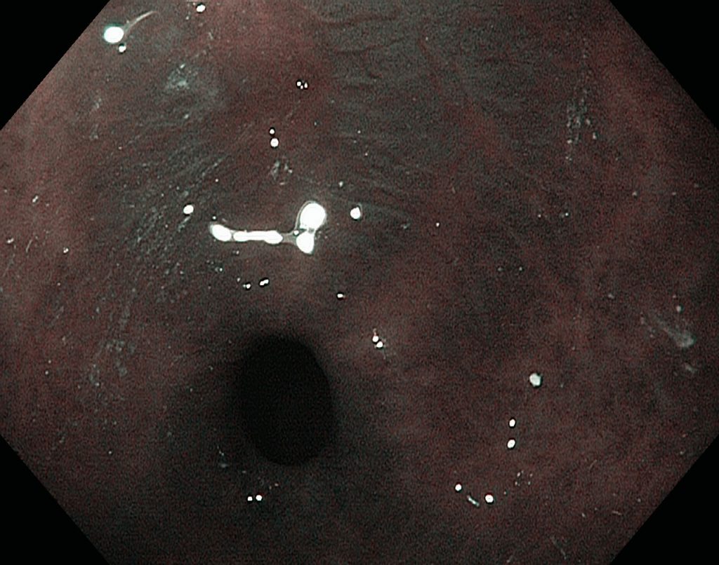
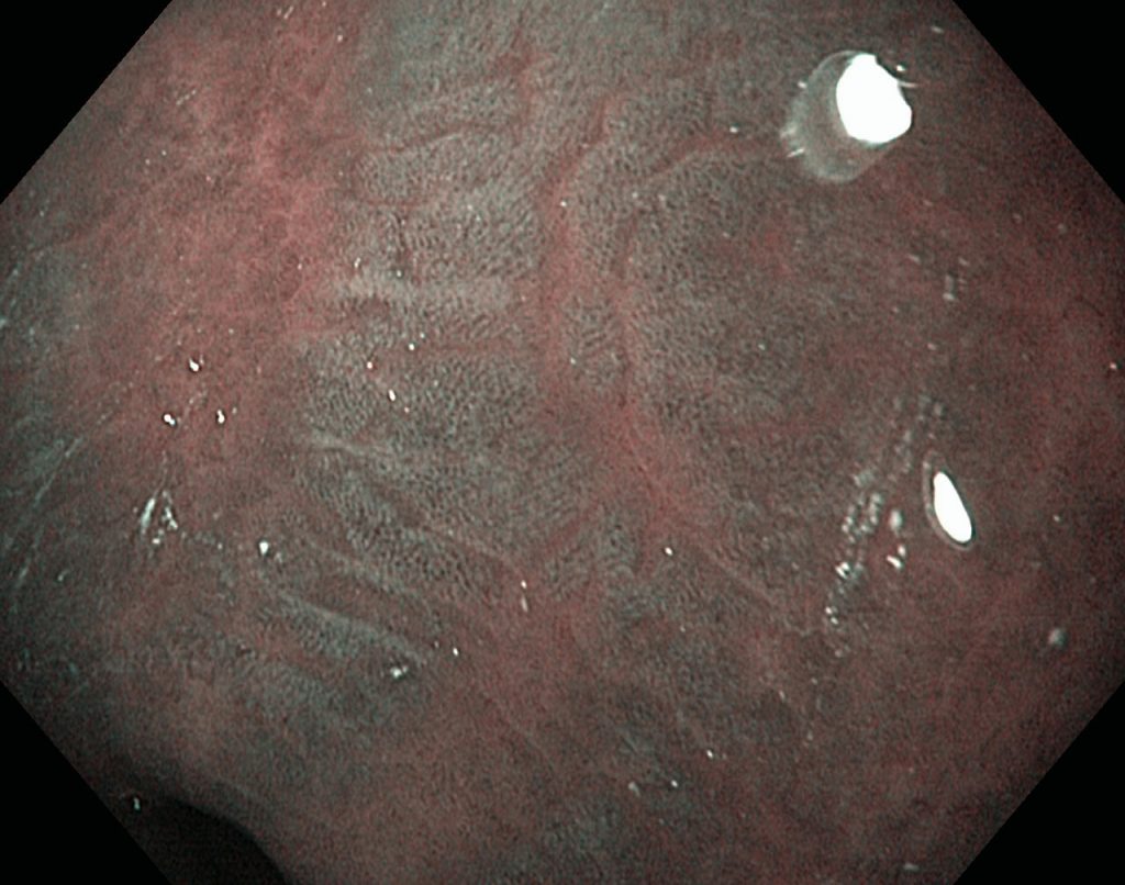
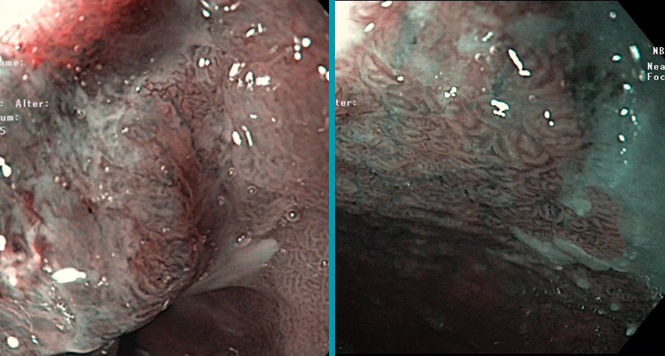
Gastric Intestinal Metaplasiais a risk factor of intestinal-type gastric cancer, but WLI was not adequate to detect IM of stomach [1]. NBI system with and without magnification can excellently diagnose GIM with good histological agreement. Uedo et al. used a light blue crest (LBC) on epithelial surface by ME-NBI as a maker to diagnose IM with the sensitivity and specificity of 89% and 93%, respectively [2]. The LBC is a fine, blue-white line on the crests of the epithelial surface, and it can only be detected under wavelengths of 400–430 nm.
- M. M. Walker, “Is intestinal metaplasia of the stomach reversible?” Gut, vol. 52, no. 1, pp. 1–4, 2003.
- N. Uedo, R. Ishihara, H. Iishi et al., “A new method of diagnosing gastric intestinal metaplasia: narrow-band imaging with magnifying endoscopy,” Endoscopy, vol. 38, no. 8, pp. 819–824, 2006.
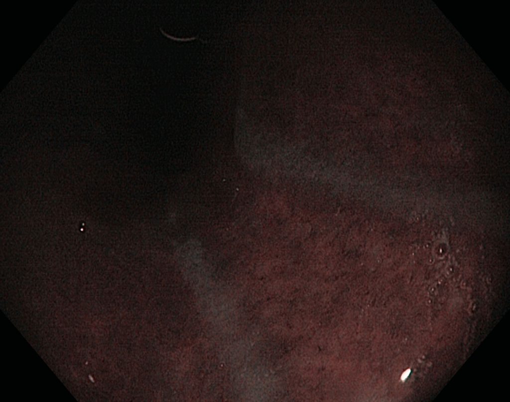
Non-magnified NBI shows the non- metaplastic mucosa as brown and the intestinal metaplasia as areas with light blue crests (LBCs)
Case A:


Case B:


Articles
Related Posts
BING Classification
Multimodal therapy for early Barrett’s neoplasias, has become established as the standard therapy and is set …

WASP classification
Recently sessile serrated lesions (SSLs) have been recognized as another important precursor lesion to CRC. SSLs …

NICE classification
The NICE (NBI International Colorectal Endoscopic) Classification is based on narrow-band images of colon polyps. The …
Our partners:
Publish Your Insights!
Share your papers, videos, articles, or classifi cations on the Endoscopy Campus and reach an international professional audience.
- International Visibility
- Professional Recognition
- Monetary Compensation
- Diverse Contribution Formats
