
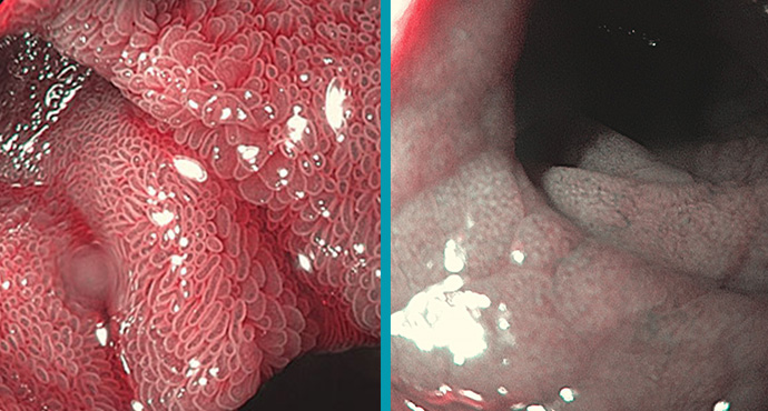
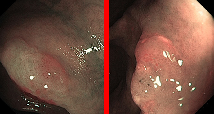
Gastric Intestinal Metaplasia is a risk factor of intestinal-type gastric cancer, but WLI was not adequate to detect IM of stomach. NBI system with and
Galleries
Galleries
Stomach: Light Blue Crest Sign for Intestinal Metaplasia
Endoscopic diagnosis of celiac disease
Celiac disease is a chronic inflammation trigged by the ingestion of gluten and resulting in a dense infiltration of lymphocytes in the proximal small intestine.

Neuroendocrine Gastric Tumors
Among the gastric submucosal tumors, neuroendocrine tumors are a special entity, which also require examination of independent gastric mucosal biopsies for classification.

Heterotopic gastric mucosa
Heterotope Magenschleimhaut des Ösophagus (heterotopic gastric mucosa, gastric inlet patch) entspricht funktionellem Magengewebe, das sich nicht an der anatomisch üblichen Lokalisation befindet. Sie ist in

When the Z-line is not completely normal
Depending on the patient’s degree of sedation and the examiner’s level of experience, carrying out a precise examination of the Z-line may not be very
Polyp Classification: WASP (incl. SSA)
The following gallery of images is intended to present the newly evaluated characteristics in the WASP classification, but it also illustrates the problems that still

Rectal NET Tumors
Unclear smaller polyps in the rectum may represent a pitfall — if they do not look like perfectly typical hyperplasia or small adenomas, then carcinoids

How can I identify sessile serrated adenomas?
Flat polyps are difficult to identify and may be easily overlooked, particularly in the right colon, where there is sometimes limited bowel cleansing. If a
