
The Forrest Classification is now used as a tool to identify patients …
Classifications
Classifications & Enitites
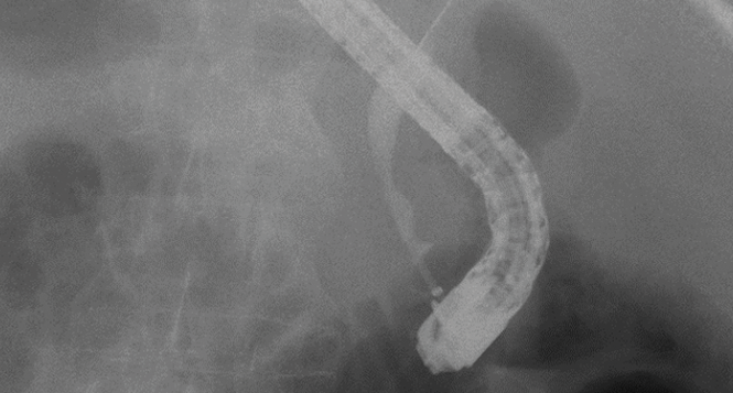
Classification and characteristics of IgG4-associated cholangitis
Classification and characteristics of IgG4-associated cholangitis
On the basis of a clinical case, typical endoscopic findings and diagnostic …
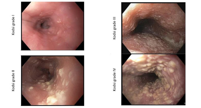
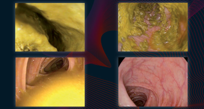
Kodsi classification of Candida esophagitis
Kodsi classification of Candida esophagitis
Candidiasis is the most frequent form of infectious esophagitis. The characteristic white …
Boston-Bowel-Preparation-Scale
Boston-Bowel-Preparation-Scale
Various measures introduced in recent decades have led to a reduction in …

Hiatus hernias and the Hill classification
Hiatus hernias and the Hill classification
To allow more precise assessment of the competence of the esophagogastric sphincter …

Follow-up intervals after polypectomy
Follow-up intervals after polypectomy
Follow-up intervals after polypectomy Colonoscopy is the most reliable procedure for detecting …

Sydney classification- assessment of deep mural injury after endoscopic mucosal resection.
Sydney classification- assessment of deep mural injury after endoscopic mucosal resection.
Classification presented by Burgess NG et al. based on retrospective evaluation, clinical …

The CAES classification of anastomotic insufficiency in the esophagus
The CAES classification of anastomotic insufficiency in the esophagus
The Surgical Working Group on Endoscopy and Ultrasound (Chirurgische Arbeitsgemeinschaft für Endoskopie …

Endosonographic Criteria Chronic Pancreatitis
Endosonographic Criteria Chronic Pancreatitis
This article provides an overview of the scoring systems commonly used for …

Tokyo Classification Cholangitis (Guidelines)
Tokyo Classification Cholangitis (Guidelines)
Acute cholangitis results from disturbed biliary drainage and bacterial infection. The mortality …

IMPN: Fukuoka Classification (Guidelines)
IMPN: Fukuoka Classification (Guidelines)
Increasing numbers of cystic tumors in the pancreas are being diagnosed. It …
BING Classification Early Barrett Neoplasia
BING Classification Early Barrett Neoplasia
Multimodal therapy for early Barrett’s neoplasias, has become established as the standard …

WASP classification – optical diagnosis of polyps <10mm
WASP classification – optical diagnosis of polyps <10mm
Recently sessile serrated lesions (SSLs) have been recognized as another important precursor …

Achalasia: Chicago Classification
Achalasia: Chicago Classification
Achalasia is one of the differential diagnoses in patients with symptoms of …

Chronic Inflammatory Bowel Disease: Endoscopic Scores
Chronic Inflammatory Bowel Disease: Endoscopic Scores
Chronic inflammatorey bowel disease (IBD) with ist two forms Crohns Disease (CD) …

Polyp Classification: BASIC
Polyp Classification: BASIC
Basic (BLI Adenoma Serrated International Classification) Classification for colorectal polyp characterization with …
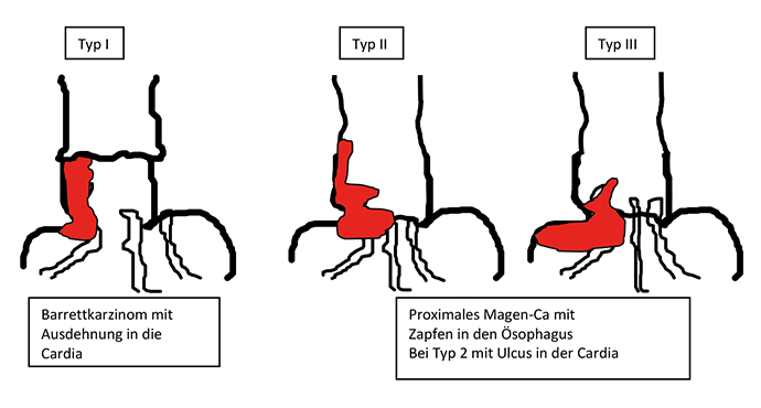
Esophagastric Junction Cancers (AEG)
Esophagastric Junction Cancers (AEG)
Tumors of the esophagogastric junction should be classified not only according to …

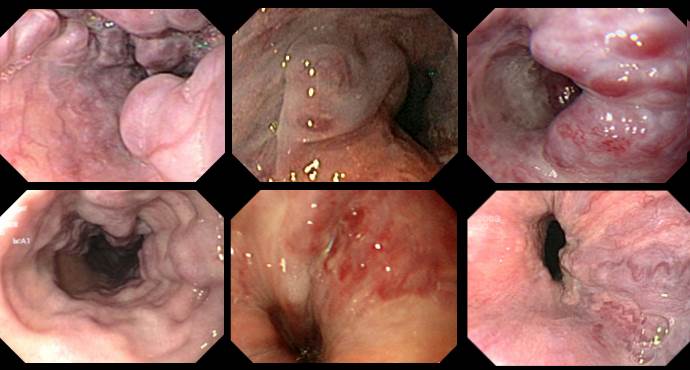
Paris Classification: Early Barrett Cancers
Paris Classification: Early Barrett Cancers
In the following, examples for superficial/early Barrett lesions of the esophagus are …

Paris Classification: Early Colorectal Cancers
Paris Classification: Early Colorectal Cancers
The Paris classification for superficial / early tumors should be part of …
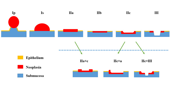
Paris Classification Early Cancer
Paris Classification Early Cancer
Endoscopic treatment for early carcinoma in the gastrointestinal tract has in the …

Paris Classification: Early Squamous Cell Cancers Esophagus
Paris Classification: Early Squamous Cell Cancers Esophagus
Examples of superficial/early squamous cell lesions in the esophagus are presented below. …

Paris Classification: Early Gastric Cancer
Paris Classification: Early Gastric Cancer
Examples of superficial/early gastric tumors are shown below. In the stomach, flat …
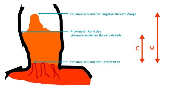
Prague Classification Barrett Esophagus
Prague Classification Barrett Esophagus
The Prague classification was presented by an international research group in 2006 …

Reflux Esophagitis: Los Angeles Classification
Reflux Esophagitis: Los Angeles Classification
Gastroesophageal reflux disease with endoscopically identifiable lesions (erosions, stricture, Barrett’s esophagus) is …

Polyp Classification: NICE
Polyp Classification: NICE
The NICE (NBI International Colorectal Endoscopic) Classification is based on narrow-band images …
Esophageal Varices
Esophageal Varices
Various systems are available for classifying esophageal varices. Unfortunately, they only overlap …

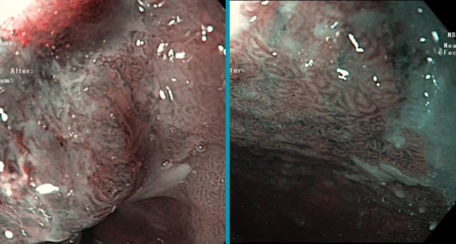
Eosinophilic Esophagitis
Eosinophilic Esophagitis
A classification for eosinophilic esophagitis has not yet been included in the …

Fundic Varices
Fundic Varices
In contrast to esophageal varices, there is only one classification system for …
