


Lars explains Anatomy – Hepaticojejunostomy
Teaching Videos
Teaching Videos
Lars explains Anatomy – Hepaticojejunostomy
Sub-cardial Gastric ESD
Before starting any endoscopical procedure, in particular an endoscopic submucosal dissection, a proper and extensive evaluation of the lesion must be accomplished. White light endoscopy,
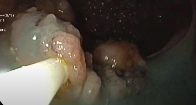
Sub-mucosal Tunneling Endoscopic Resection (STER) of an Esophageal Leiomyoma
A two centimeter submucosal lesion arising from muscularis mucosae is identified in the distal esophagus.
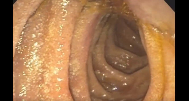
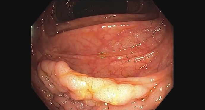
Piecemeal endoscopic mucosal resection (EMR) of an extensive laterally spreading adenoma in the rectum
Piecemeal endoscopic mucosal resection (EMR) of an extensive laterally spreading adenoma in the rectum

Z-POEM -a combination of septomoty and tunnel myotomy
Z-POEM -a combination of septomoty and tunnel myotomy

Rectal Endoscopic Submucosal Dissection
In this case, a 65-year-old woman presents with a recurrence of a polyp previously treated with endoscopic mucosal resection. The biopsy and the endoscopic evaluation

Pediatric Per Oral Endoscopic Myotomy (Posterior Approach)
This video presents the case of a five-year-old child with confirmed type two achalasia. For POEM, patient is positioned in supine and insufflation with CO2

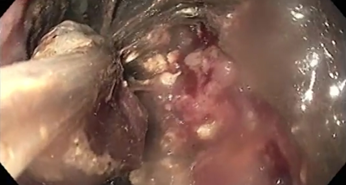
Recovery of a lost gallstone from a perihepatic abscess
Gallbladder perforation, with stone “emptying” into the abdominal cavity, is a potential complication during cholecystectomy. Later complications may develop, particularly abscess formation. A subphrenic abscess
Endoscopic removal of a buried bumper using the Flamingo system
A patient with an ingrown internal PEG retaining plate (buried bumper syndrome) on the anterior wall of the gastric body. Endoscopic removal of the device
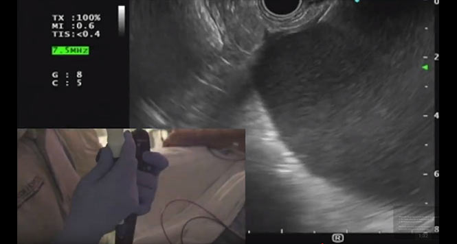
A Case with an Abdominal Fluid Collection
This video demonstrates the case of a 42-year old male referred for EUS-guided drainage of a pancreatic fluid collection.
Acute necrotic collection
This video demonstrates the features of an acute necrotic collection in the case of a 65-year old male with abdominal pain and fever.
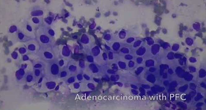
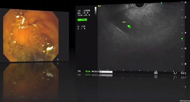
Cancer within a Pancreatic Fluid Collection
This video demonstrates EUS-guided drainage of a pancreatic fluid collection, and illustrates the importance of a careful EUS examination in all patients referred for PFC
Challenges of EUS-FNA of an Uncinate Pancreatic Mass
This video demonstrates the challenges associated with EUS-guided FNA of a pancreatic uncinate mass.

Duplication Cyst vs. Spindle Cell Tumor on EUS
This video demonstrates the differences between duplications cysts and spindle cell tumors.

EUS Features of Disconnected Pancreatic Duct Syndrome
This video demonstrates the hallmarks of Disconnected Pancreatic Duct Syndrome.

EUS-FNA of a Pancreatic Uncinate Mass using the Fanning Technique
This video demonstrates the EUS-FNA of a pancreatic uncinate mass using the Fanning Technique.

EUS-Guided Celiac Plexus Neurolysis
This video demonstrates the technique of EUS-guided Celiac Plexus Neurolysis in a patient with unresectable pancreatic adenocarcinoma.

EUS-Guided drainage of PFC using a Lumen-Apposing Metal Stent: Single-Gate Technique
This video demonstrates the single-gate technique of drainage of a pancreatic fluid collection using a lumen-apposing metal stent.

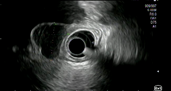
The importance of water insufflation in the EUS evaluation of a subepithelial lesion
This video demonstrates the importance of water insufflation in the EUS evaluation of a subepithelial lesion seen in the duodenal bulb on EGD, in the

Establishment of an endoscopic gastroenterostomy
In this video Dr. Wannhoff from Ludwigsburg shows a new, minimally invasive approach to the endosonographic attachment of a gastroenterostomy in gastric emptying disorder.

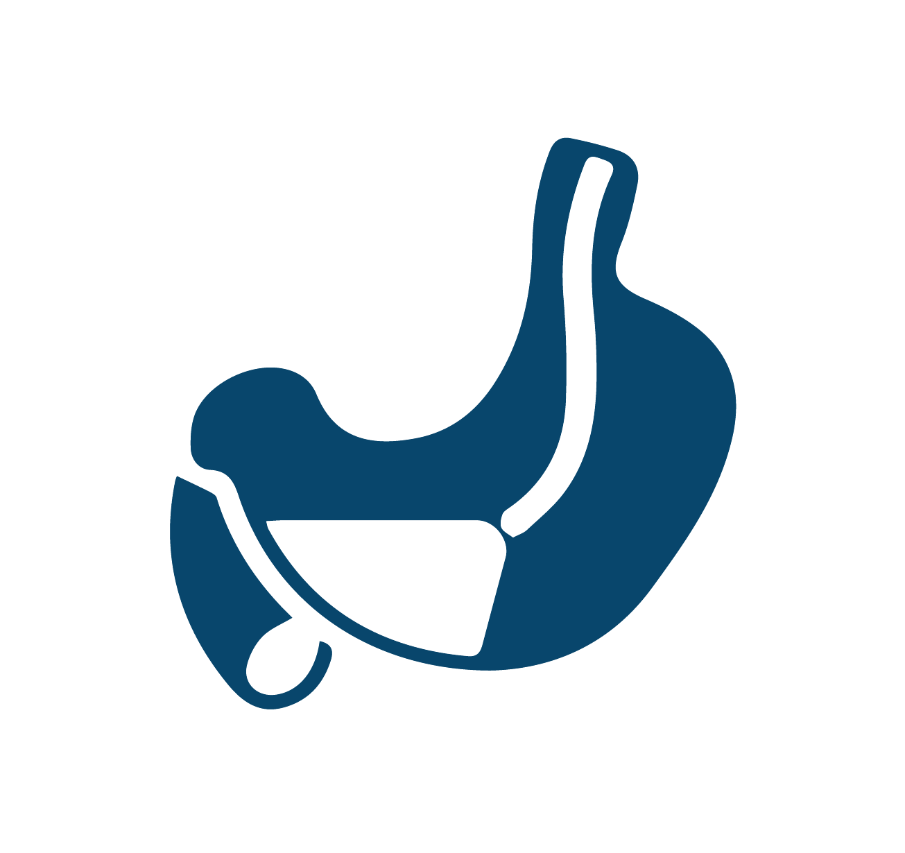
Lars explains Anatomy – Gastric Bypass
Die Anzahl an durchgeführten Adipositas-OPs steigt weltweit. Für jeden Endoskopiker ist es wichtig die Anatomie nach einer Magen-Bypass-OP zu kennen um evtl. Komplikationen nach diesen


Endoscopic relief of a gallbladder empyema
The endoscopic ultrasound (EUS) image, seen from the antrum, shows a tightly filled gallbladder with hyperechoic reflexes, in a 71-year-old patient. The patient had an

The bougie cap – a new method of treating stenoses in the gastrointestinal tract
In the classic method, a stricture in the esophagus is dilated using a Savary bougie after advancement of a guide wire. The difficulty with this

Roux-en-Y anatomy after gastric resection
This video illustrates the altered anatomy resulting after the type of gastric resection that is carried out for gastric carcinoma, for example. Bowel continuity is
Multimodal treatment approach with endoscopic full-thickness resection of a rectal carcinoma
A 77-year-old patient presented to the emergency department with Hb-relevant lower gastrointestinal bleeding during anticoagulation treatment with rivaroxaban and clopidogrel. At colonoscopy, the bleeding source
Endoscopic full-thickness resection of a GIST using GERD-X
A subepithelial tumor has been identified in the fundus. EUS shows that it is 2.5 × 3 cm in size, probably arising from the muscularis propria. No pathological

Billroth II anatomy after partial stomach resection
This video explains the altered anatomy that is encountered after a Billroth II operation. In a Billroth II resection, the lower part of the stomach

Gastric peroral endoscopic myotomy (G-POEM)
Hendrik Manner from Wiesbaden reports on a patient with a gastric emptying disorder who was treated with what is known as gastric peroral endoscopic myotomy

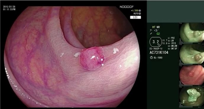
Distal hypoechoic submucosal tumor in the esophagus
Submucosal lesions identified in the esophagus usually undergo further clarification using endoscopic ultrasonography (EUS). In this video, Thomas Rösch from Hamburg demonstrates the examination sequence
Distaler echoarmer submuköser Tumor im Ösophagus
Submuköse Läsionen im Ösophagus werden endoskopisch meist als Zufallbefund diagnostiziert und im Weiteren mittels Endosonographie abgeklärt. Thomas Rösch aus Hamburg zeigt in diesem Video den
The EndoRotor® as a completely new mechanical mucosectomy procedure — an alternative for faster ER and ESD?
Stephan Hollerbach and his team demonstrate an en-bloc resection in a swine model using the new mechanical EndoRotor® resection system.
Transgastric biliary drainage with metastatic gallbladder cancer
Various access routes are available for drainage in patients with cholestasis. In the complex case presented here, neither ERCP nor an attempt at PTCD placement

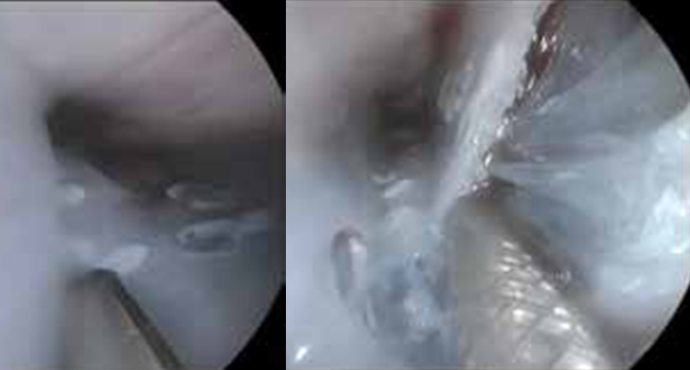
Tunnel removal of a submucosal tumor in the esophagus (SET technique)
Dr. Werner and Prof. Rösch from Hamburg present the case of a young patient with an incidental finding of esophageal GIST. In this patient, it

Endocuff Vision® versus wide-angle endoscope: small attachment – big effect
The wide-angle colonoscope provides a field of view up to 235 degrees, thanks to a lens system featuring forward viewing (147 degrees) and also side
Endoscopic therapy of pancreatic fluid collections caused by severe necrotic pancreatitis
Pancreatitis can cause various severe complications such as acute fluid collections with superinfected necrotic content requiring drainage and removal of necrotic debris. Here we demonstrate
Early esophageal carcinoma (squamous epithelium): tips and tricks for difficult ESDs
Early esophageal carcinoma (squamous epithelium): tips and tricks for difficult ESDs

Post-EMR arterial bleeding
Arterial bleeding from the area of the endoscopic mucosal resection, 2 days after the intervention. Successful hemostasis is achieved using bipolar coagulation forceps in “Soft

Treatment for papillary adenoma
Treatment for papillary adenoma. An injection is made into the papillary adenoma to produce a good lifting sign.

PEXACT — direct-puncture PEG after gastropexy
The gastropexy device consists of two hollow needles that are attached to each other. A suture thread is inserted through one hollow needle, and a

Endoscopic division of a Zenker diverticulum using the Clutch Cutter
Endoscopic division of a Zenker diverticulum using the Clutch Cutter and management of a perforation. Coagulation of the diverticular septum using the Clutch Cutter. Settings:
Endoscopic diagnosis of colon polyps using the NICE classification: initial experience with BLI and LCI
The new generation of the Fujifilm electronic endoscopy system allows virtual chromoendoscopy alongside zoom endoscopy. Blue laser imaging (BLI), like narrow-band imaging (NBI), allows further

Small carcinoma in Barrett’s esophagus — EMR and RFA
A 46-year-old patient with short-segment Barrett’s esophagus that had been receiving monitoring since 2009, now presenting with a mucosal adenocarcinoma.
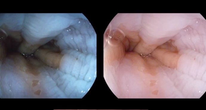
Endoscopic examination of a normal Z-line
Visualization of the Z-line without enhancement and with iScan, obstructed by esophageal motility.

Perforation of a bleeding polypectomy site in the colon using a Hemoclip
A patient who had undergone polypectomy of a flat polyp in the ascending colon presented again with peranal hemorrhage. The polypectomy site can be seen

Combined laparoscopic/endoscopic resection of a cecal adenoma
A 60-year-old female patient with a diagnosis of a cecal adenoma that cannot be resected endoscopically, developing out of the appendix.

Endoscopy antireflux therapy with the MUSE system
A 31-year-old female patient who has had reflux symptoms for 15 years and has responded well to PPI therapy. The patient wants to stop taking
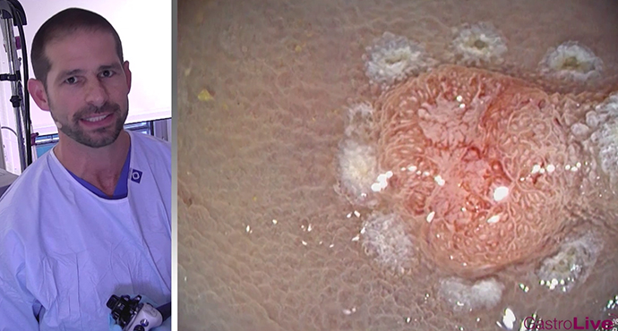
Piecemeal EMR of a laterally spreading tumor (LST)
Diagnosis of a granular-type laterally spreading tumor at the right colic flexure.
