
The Forrest Classification is now used as a tool to identify patients who are at an increased risk for bleeding, rebleeding and mortality
Classifications
Classifications
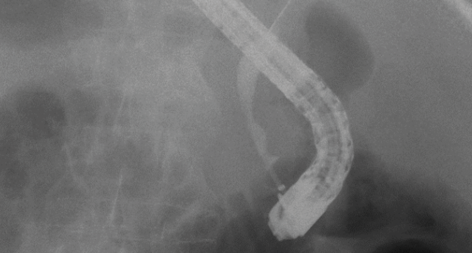
Classification and characteristics of IgG4-associated cholangitis
Classification and characteristics of IgG4-associated cholangitis
On the basis of a clinical case, typical endoscopic findings and diagnostic criteria for hepatobiliary IgG4 disease are described here, along with the classification of
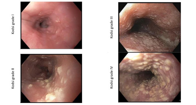
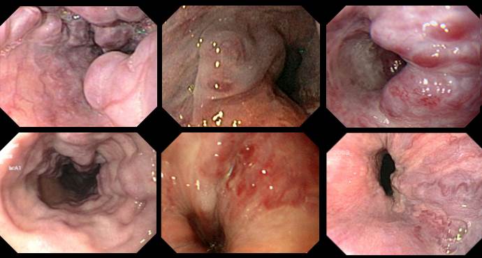
Kodsi classification of Candida esophagitis
Kodsi classification of Candida esophagitis
Candidiasis is the most frequent form of infectious esophagitis. The characteristic white plaques, which are difficult to rinse off, are found in approximately 4% of
Boston-Bowel-Preparation-Scale
Boston-Bowel-Preparation-Scale
Various measures introduced in recent decades have led to a reduction in the mortality rate due to colorectal carcinoma. They include screening colonoscopy.

Hiatus hernias and the Hill classification
Hiatus hernias and the Hill classification
To allow more precise assessment of the competence of the esophagogastric sphincter mechanism, Hill et al. presented a practical classification in 1996.

Follow-up intervals after polypectomy
Follow-up intervals after polypectomy
Follow-up intervals after polypectomy Colonoscopy is the most reliable procedure for detecting colorectal carcinomas and polyps. The aim of colonoscopy must be to achieve a

Sydney classification- assessment of deep mural injury after endoscopic mucosal resection.
Sydney classification- assessment of deep mural injury after endoscopic mucosal resection.
Classification presented by Burgess NG et al. based on retrospective evaluation, clinical observations and image analysis. It allows for the assessment of deep mural injury

The CAES classification of anastomotic insufficiency in the esophagus
The CAES classification of anastomotic insufficiency in the esophagus
The Surgical Working Group on Endoscopy and Ultrasound (Chirurgische Arbeitsgemeinschaft für Endoskopie und Sonographie, CAES) has developed and validated a classification of anastomotic insufficiency in

Endosonographic Criteria Chronic Pancreatitis
Endosonographic Criteria Chronic Pancreatitis
This article provides an overview of the scoring systems commonly used for diagnosing chronic pancreatitis by means of endoscopic ultrasonography (EUS), and for classifying the

Tokyo Classification Cholangitis (Guidelines)
Tokyo Classification Cholangitis (Guidelines)
Acute cholangitis results from disturbed biliary drainage and bacterial infection. The mortality rates due to acute cholangitis reported in the literature over the last 20

IMPN: Fukuoka Classification (Guidelines)
IMPN: Fukuoka Classification (Guidelines)
Increasing numbers of cystic tumors in the pancreas are being diagnosed. It is often difficult to precisely assign these highly varied tumors to a specific
BING Classification Early Barrett Neoplasia
BING Classification Early Barrett Neoplasia
Multimodal therapy for early Barrett’s neoplasias, has become established as the standard therapy and is set out in national and international guidelines. These dysplastic lesions
Endosonographic Criteria Chronic Pancreatitis
Endosonographic Criteria Chronic Pancreatitis
This article provides an overview of the scoring systems commonly used for diagnosing chronic pancreatitis by means of endoscopic ultrasonography (EUS), and for classifying the

WASP classification – optical diagnosis of polyps <10mm
WASP classification – optical diagnosis of polyps <10mm
Recently sessile serrated lesions (SSLs) have been recognized as another important precursor lesion to CRC. SSLs are thought be responsible for 15–30% of colorectal cancer.

Achalasia: Chicago Classification
Achalasia: Chicago Classification
Achalasia is one of the differential diagnoses in patients with symptoms of dysphagia. High-resolution (HR) manometry is now regarded as the diagnostic gold standard for

Chronic Inflammatory Bowel Disease: Endoscopic Scores
Chronic Inflammatory Bowel Disease: Endoscopic Scores
Chronic inflammatorey bowel disease (IBD) with ist two forms Crohns Disease (CD) and Ulcerative Colitis (UC) can be classified by various endoscopic scores with regards

Polyp Classification: BASIC
Polyp Classification: BASIC
Basic (BLI Adenoma Serrated International Classification) Classification for colorectal polyp characterization with blue light imaging
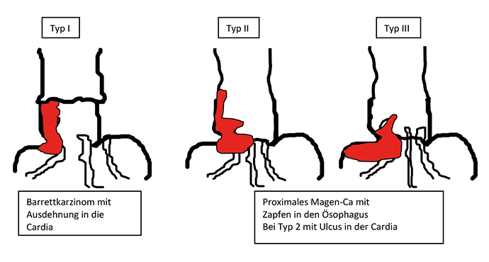
Esophagastric Junction Cancers (AEG)
Esophagastric Junction Cancers (AEG)
Tumors of the esophagogastric junction should be classified not only according to the TNM system with regards to tumor penetration (T stage), presence of lymph

Paris Classification: Early Barrett Cancers
Paris Classification: Early Barrett Cancers
In the following, examples for superficial/early Barrett lesions of the esophagus are shown. Here, flat and sessilelesions are predominant, pedunculated tumors are rare. Sessile tumor

Paris Classification: Early Colorectal Cancers
Paris Classification: Early Colorectal Cancers
The Paris classification for superficial / early tumors should be part of a standard terminology for endoscopic assessment. This classification applies to the entire gastrointestinal
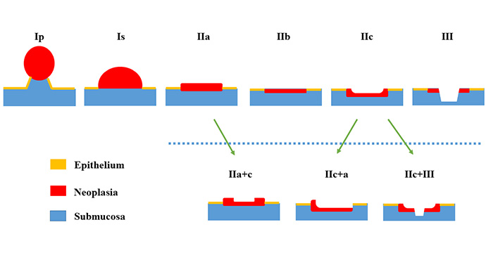
Paris Classification Early Cancer
Paris Classification Early Cancer
Endoscopic treatment for early carcinoma in the gastrointestinal tract has in the meantime become evidence-based and has been incorporated into national and international guidelines

Paris Classification: Early Squamous Cell Cancers Esophagus
Paris Classification: Early Squamous Cell Cancers Esophagus
Examples of superficial/early squamous cell lesions in the esophagus are presented below. In the esophagus, flat lesions are predominant in the early tumors, and polypoid

Paris Classification: Early Gastric Cancer
Paris Classification: Early Gastric Cancer
Examples of superficial/early gastric tumors are shown below. In the stomach, flat lesions are predominant, often as combined lesions with a central depression (IIa+c). Sessile
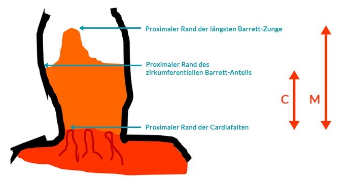
Prague Classification Barrett Esophagus
Prague Classification Barrett Esophagus
The Prague classification was presented by an international research group in 2006 (1) and has since been regarded as the standard for measuring the length

Reflux Esophagitis: Los Angeles Classification
Reflux Esophagitis: Los Angeles Classification
Gastroesophageal reflux disease with endoscopically identifiable lesions (erosions, stricture, Barrett’s esophagus) is defined as erosive gastroesophageal reflux disease (GERD). Fewer than 50% of patients with

Polyp Classification: NICE
Polyp Classification: NICE
The NICE (NBI International Colorectal Endoscopic) Classification is based on narrow-band images of colon polyps. The classification uses staining, vascular patterns, and surface patterns to
Esophageal Varices
Esophageal Varices
Various systems are available for classifying esophageal varices. Unfortunately, they only overlap or coincide partly. The official terminology used by the German Society for Digestive

Eosinophilic Esophagitis
Eosinophilic Esophagitis
A classification for eosinophilic esophagitis has not yet been included in the usual terminologies in the German-speaking countries. In the official terminology of the German

Fundic Varices
Fundic Varices
In contrast to esophageal varices, there is only one classification system for fundic varices, developed by Sarin et al.
