Paris Classification: Early Squamous Cell Cancers Esophagus
Thomas Rösch, Hamburg
The Paris classification of superficial tumors, which is presented and discussed in detail in the introductory section, should be used in standard endoscopic terminology. A diagram of the classification is shown again below:

Examples of superficial/early squamous cell lesions in the esophagus are presented below. In the esophagus, flat lesions are predominant in the early tumors, and polypoid and pedunculated tumors are very rare. Sessile tumors with substantial intraluminal growth are often no longer early cancers; the same also applies to excavated lesions of various sizes (type IIc or even III).
Examples of squamous cell tumors, 1: type I, elevated


The lesion on the left is centrally less elevated/slightly depressed. Localized elevated squamous cell cancers as shown in the right image are rare. Histology in both cases: T1sm.
Examples of squamous cell tumors, 2: type IIa, flat and elevated

Histology: Biopsy at an external institution: high-grade dysplasia; histology after EMR: T1 m, basal R0.
Examples of squamous cell tumors, 3: type IIb, flat


Histology: Biopsy at an external institution: high-grade dysplasia; histology after EMR: T1 sm, basal R0; after surgery: no residual tumor in the esophagus, but N1 (1/18 LN)


Histology: only HGIN.


The position of the endoscope differed in the two pictures, and the right image has therefore been rotated to match the left one. Histology: T1m.
Examples of squamous cell tumors, 4: type IIb, depressed
With these types, there is a kind of slightly elevated margin, and many of these lesions could therefore also be classified as type IIa+c.


Related Posts
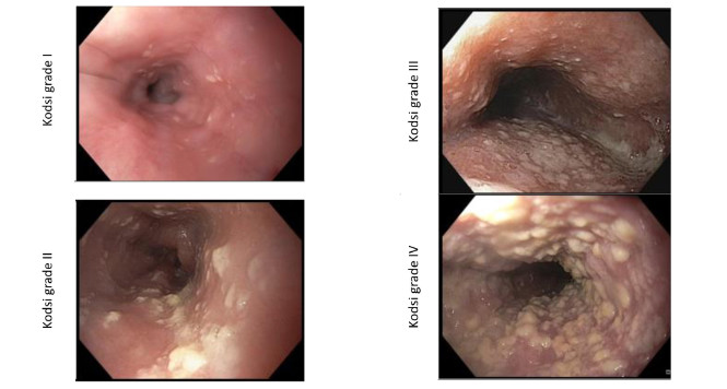
Kodsi classification of Candida esophagitis
Candidiasis is the most frequent form of infectious esophagitis. The
KLASSIFIKATION ANSEHEN
STER for Esophageal Leiomyoma
A two centimeter submucosal lesion arising from muscularis mucosae is
WATCH THE VIDEO