Fundic Varices
Sarin classification
- Gastroesophageal varices, type 1 (on the endoscopic appearance, a continuation of esophageal varices into the lesser curvature of the stomach beyond the level of the cardia, usually with an elongated course)
- Gastroesophageal varices, type 2 (endoscopically, pass from the cardia towards the greater curvature into the fundus of the stomach, often with a sinuous, cluster-like course)
- • Isolated gastric varices, type 1 (this is the term for varices that course in the gastric fundus or cardia, but do not pass into the esophagus and do not reach the cardia)
- • Isolated gastric varices, type 2 (this is the term for “ectopic” varices in all other sections of the stomach)
The only other parameter used is the maximum size of the varix in millimeters.
For classification of varices in the fundus, the esophagogastric junction should be viewed in a prograde fashion, and the fundus and cardia should then be viewed in inversion with good insufflation.
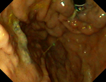
Examples of type 1 gastroesophageal varices
Extension of varices across the cardia toward the lesser curvature and into the gastric fundus.
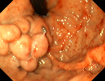
Examples of type 2 gastroesophageal varices
Extension of varices beyond the cardia toward the greater curvature and into the gastric fundus.
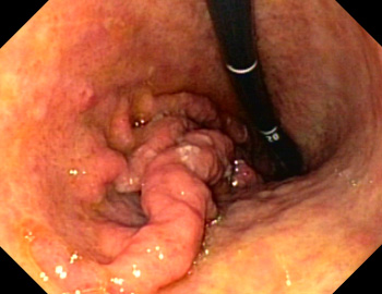
Examples of type 1 isolated gastric varices
The varices course in the gastric fundus or cardia, but do not pass into the esophagus and do not reach the cardia.
Examples of type 2 isolated gastric varices
“Ectopic” varices in all other sections of the stomach (below the fundus), in this case in the gastric body.
General notes on the classifications
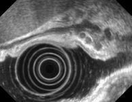
Classifications of varices apply strictly only to the primary assessment, before (endoscopic) therapy; classifications after treatment (injection therapy) are more difficult. In that case, the (residual) varices should be described in terms of number, location, and maximum diameter or red signs. On endoscopic ultrasound (EUS), smaller vessels are often seen in the walls, but these are of no relevance. However, EUS can demonstrate residual flow when the endoscopic findings are unclear after therapy.
Example images
Endoscopic image of a fundic varix after Histoacryl injection. On EUS, a fully lined varix with hyperechoic areas (Histoacryl) is seen, with an acoustic shadow, and with small intramural wall varices alongside that are of no clinical relevance.
There have been no studies directly dealing with the issue of interobserver variability in classifying fundic varices, only on the general assessment of gastric signs of portal hypertension [3, 4].
References
- Sarin SK, Kumar A. Gastric varices: profile, classification, and management. Am J Gastroenterol. 1989;84(:1244-9.
- Sarin SK, Lahoti D, Saxena SP, Murthy NS, Makwana UK. Prevalence, classification and natural history of gastric varices: a long-term follow-up study in 568 portal hypertension patients. Hepatology. 1992;16:1343-9.
- Calès P, Buscail L, Bretagne JF, Champigneulle B, Bourbon P, Duclos B, Dapoigny M, Dumas R, Pierrugues R, Davion T, et al. Interobserver and intercenter agreement of gastro-esophageal endoscopic signs in cirrhosis. Results of a prospective multicenter study Gastroenterol Clin Biol. 1989 Dec;13(12):967-73.
- Calès P, Zabotto B, Meskens C, Caucanas JP, Vinel JP, Desmorat H, Fermanian J, Pascal JP. Gastroesophageal endoscopic features in cirrhosis. Observer variability, interassociations, and relationship to hepatic dysfunction. Gastroenterology. 1990;98:156-62.


