Kodsi classification of Candida esophagitis
Dr. med. Daniel Fitting
Universitätsklinikum Würzburg Medizinische Klinik und Poliklinik II
Candidiasis is the most frequent form of infectious esophagitis. The characteristic white plaques, which are difficult to rinse off, are found in approximately 4% of patients who undergo esophagogastroduodenoscopy (EGD) [1]. The plaques consist of leukocytes, necrotic mucosa and Candida pseudohyphae. The most frequent pathogen is C. albicans, with C. glabrata and C. krusei being only rarely found — so that further microbiological diagnosis is not usually required before local treatment with amphotericin B or systemic triazole therapy.
The disease becomes clinically manifest with dysphagia, epigastric pain, or oral candidiasis. Forty percent of the patients remain asymptomatic [2]. Risk factors for Candida esophagitis include immunosuppression and motility disturbances in the esophagus. However, temporary factors such as proton pump inhibitor (PPI) treatment, local steroids, antibiotic therapy, poorly controlled diabetes and malnutrition also favor local invasion by the yeastlike fungi, which are found in the normal gastrointestinal flora [3].
When there is mild involvement in the upper third of the esophagus, local therapy with amphotericin B or nystatin may be considered. However, esophageal involvement usually requires systemic therapy, which should be carried out using fluconazole 200–400 mg/day orally for 14–21 days. If the patient has marked dysphagia, treatment may have to be started parenterally with fluconazole 6 mg/kg body weight per day. Itraconazole/voriconazole or echinocandins are available for refractory courses [4].
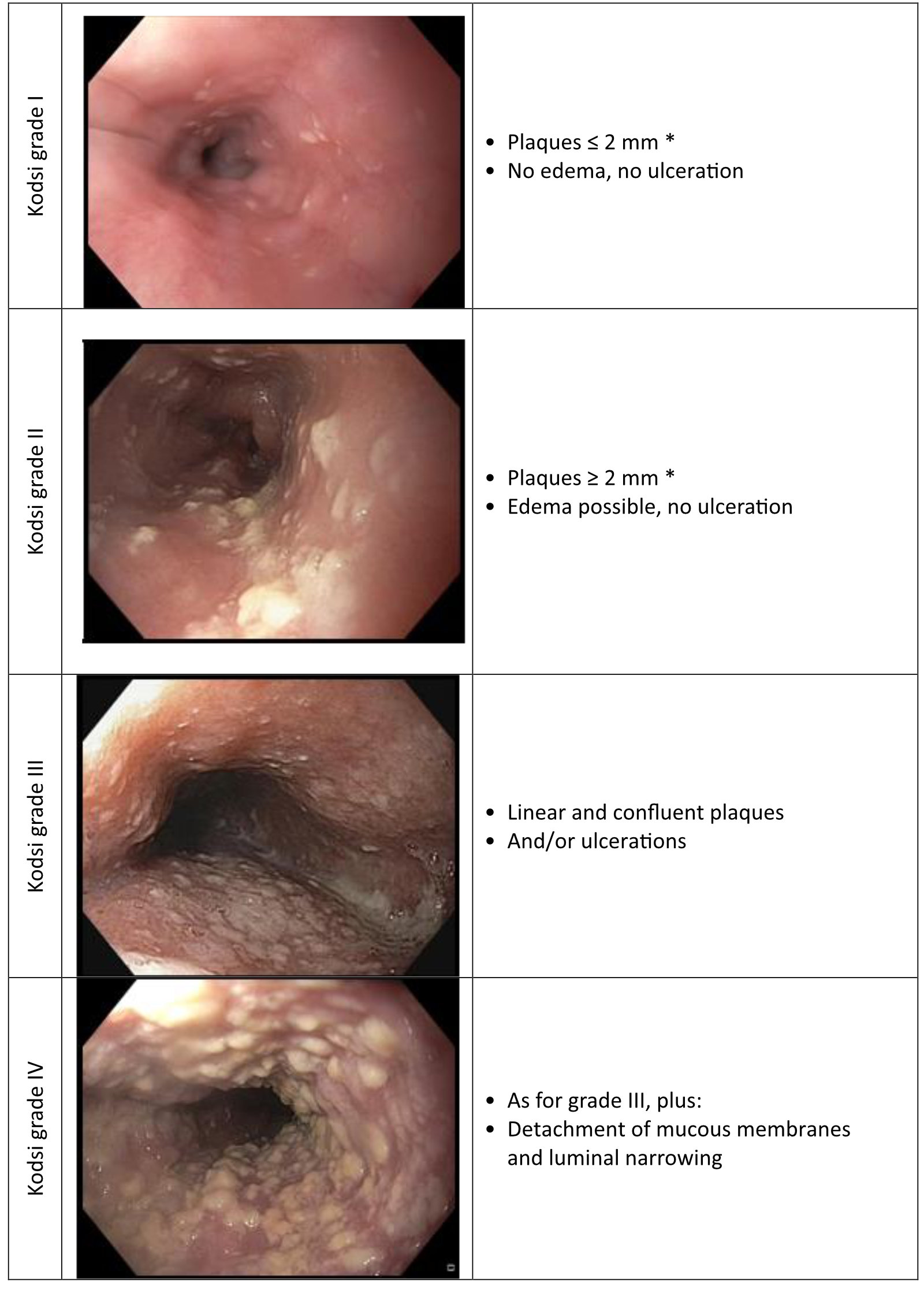
The extent of esophageal involvement with Candida can be assessed endoscopically using the Kodsi classification (Fig. 1) [2]. This correlates with the extent of the clinical symptoms and the CD4 cell count in patients with HIV [1]. The classification is easily remembered and allows structured diagnostic assessment at a high academic level.
Fig. 1 The Kodsi classification
1. Asayama N, Nagata N, Shimbo T, et al. Relationship between clinical factors and severity of esophageal candidiasis according to Kodsi’s classification. Dis Esophagus 2014; 27: 214–219.
2. Kodsi BE, Wickremesinghe C, Kozinn PJ, et al. Candida esophagitis: a prospective study of 27 cases. Gastroenterology 1976; 71: 715–719.
3. Mushi MF, Ngeta N, Mirambo MM, et al. Predictors of esophageal candidiasis among patients attending endoscopy unit in a tertiary hospital, Tanzania: a retrospective cross-sectional study. Afr Health Sci 2018; 18: 66–71.
4. Pappas PG, Kauffman CA, Andes DR, et al. Clinical Practice Guideline for the Management of Candidiasis: 2016 Update by the Infectious Diseases Society of America. Clin Infect Dis 2016; 62: e1-50.


