WASP classification – optical diagnosis of polyps <10mm
A.G.C. Bleijenberg, Amsterdam
Historically, polyps were divided into mainly adenomas and hyperplastic polyps (HPs). Adenomas were thought to be the sole precursor lesion to colorectal cancer (CRC), whereas HPs were considered harmless. Optical diagnosis strategies have therefore focused on differentiation between adenomas and HPs. For example, the Kudo, NBI International Endoscopic (NICE) and Japanese NBI Expert Team (JNET) classifications are very useful and effective for differentiation between adenomatous and non-adenomatous polyps.1–3
Recently, however, sessile serrated lesions (SSLs) have been recognized as another important precursor lesion to CRC. SSLs are thought be responsible for 15–30% of colorectal cancer.4, 5 SSL is also often referred to as sessile serrated polyp (SSP), sessile serrated adenoma (SSA) or SSA/P.
Unfortunately, SSLs are often diagnosed as non-neoplastic using the JNET or NICE classifications.3, 6 New optical diagnosis strategies have therefore been developed to include identification of SSLs as well.
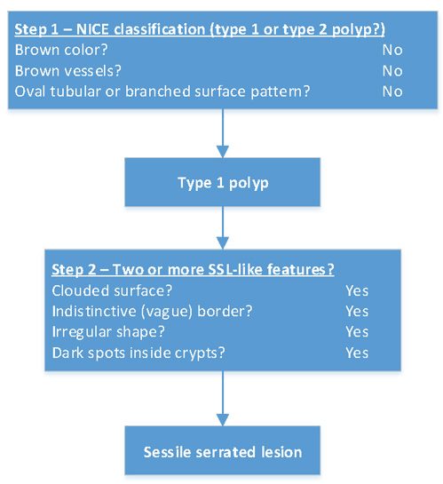
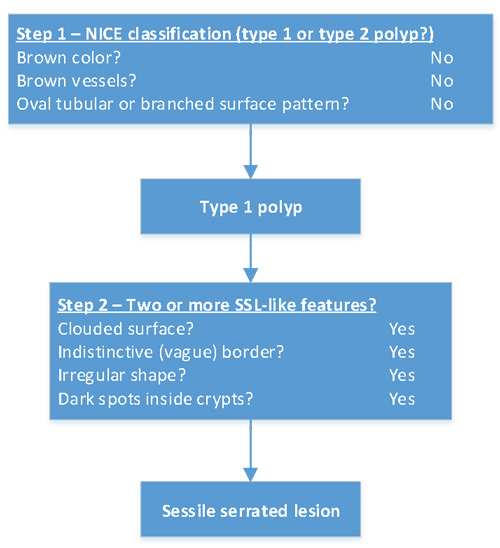
One such classification method is the WASP classification.7 The WASP aims to differentiate between 1) Hyperplastic polyps; 2) Sessile serrated lesions and 3) conventional adenomas. It uses the NICE classification2 as a first step to differentiate between type 1 and type 2 polyps. In the second step, the presence of several ‘SSL-like features’ is assessed.8 These SSL-like features are unique to SSLs, and are less often seen in adenomas or HPs. Figure 1 shows the step-wise classification.

Step 1
Differentiation between NICE type 1 and NICE type 2 polyps
Characteristic 1: Color
Same or lighter than background
Browner relative to background
Characteristic 2: Vessels
None, or isolated lacy vessels
Brown vessels surrounding white structures
Characteristic 3: Surface pattern
Dark or white spots of uniform size, or homogenous absence of pattern.
Oval, tubular or branched white structures surrounded by brown vessels
Step 2
Differentiation between hyperplastic polyp, sessile serrated lesion and adenoma based on the presence of SSL-like features
Characteristic 1 – Surface
Cloud-like surface
Smooth surface
Smooth surface
Characteristic 2 – Border
Characteristic 3 – Shape
Irregular shape
Symmetric shape
Symmetric shape
Characteristic 4 – Crypts
Dark spots inside the crypts
No dark spots inside crypts
No dark spots inside crypts
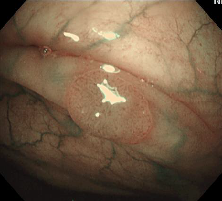
Example 1
Example 2
Example 3
Example 4
Example 5
These images are property of the Academic University Medical Centers Amsterdam, AMC, department of Gastroenterology and hepatology. Not to be reproduced without permission.
Literatur:
- Kudo S, Tamura S, Nakajima T, et al. Diagnosis of colorectal tumorous lesions by magnifying endoscopy. Gastrointest Endosc 1996;44:8-14.
- Hewett DG, Kaltenbach T, Sano Y, et al. Validation of a simple classification system for endoscopic diagnosis of small colorectal polyps using narrow-band imaging. Gastroenterology 2012;143:599-607.e1.
- Sano W, Sano Y, Iwatate M, et al. Prospective evaluation of the proportion of sessile serrated adenoma/polyps in endoscopically diagnosed colorectal polyps with hyperplastic features. Endosc Int Open 2015;3:E354-8.
- Jass JR. Classification of colorectal cancer based on correlation of clinical, morphological and molecular features. Histopathology 2007;50:113-130.
- Rex DK, Ahnen DJ, Baron JA, et al. Serrated lesions of the colorectum: review and recommendations from an expert panel. The American journal of gastroenterology 2012;107:1315-29; quiz 1314, 1330.
- Sumimoto K, Tanaka S, Shigita K, et al. The diagnostic performance of JNET classification for differentiation among noninvasive, superficially invasive, and deeply invasive colorectal neoplasia. Gastrointest Endosc 2017.
- Ijspeert JEG, Bastiaansen BaJ, van Leerdam ME, et al. Development and validation of the WASP classification system for optical diagnosis of adenomas, hyperplastic polyps and sessile serrated adenomas/polyps. Gut 2016;65:963-70.
- Hazewinkel Y, López-Cerón M, East JE, et al. Endoscopic features of sessile serrated adenomas: Validation by international experts using high-resolution white-light endoscopy and narrow-band imaging. Gastrointestinal Endoscopy 2013;77:916-924.


