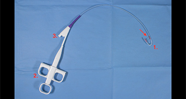
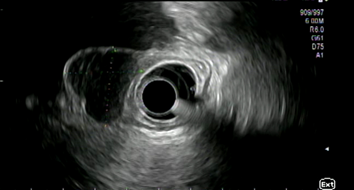
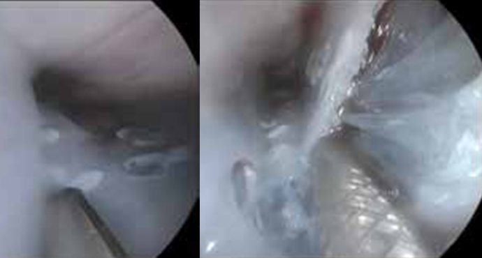
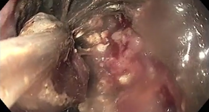
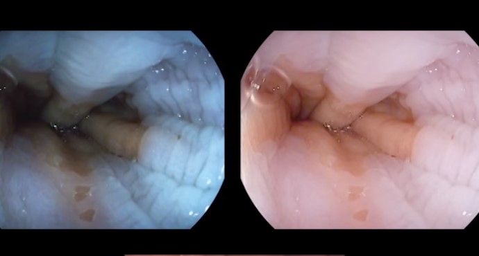
A patient with an ingrown internal PEG retaining plate (buried bumper syndrome) on the anterior wall of the gastric body. Endoscopic removal of the device
Upper GI Tract
Upper GI Tract
Endoscopic removal of a buried bumper using the Flamingo system
Lars explains Anatomy – Gastric Bypass
Die Anzahl an durchgeführten Adipositas-OPs steigt weltweit. Für jeden Endoskopiker ist es wichtig die Anatomie nach einer Magen-Bypass-OP zu kennen um evtl. Komplikationen nach diesen
The bougie cap – a new method of treating stenoses in the gastrointestinal tract
In the classic method, a stricture in the esophagus is dilated using a Savary bougie after advancement of a guide wire. The difficulty with this
Roux-en-Y anatomy after gastric resection
This video illustrates the altered anatomy resulting after the type of gastric resection that is carried out for gastric carcinoma, for example. Bowel continuity is
Endoscopic full-thickness resection of a GIST using GERD-X
A subepithelial tumor has been identified in the fundus. EUS shows that it is 2.5 × 3 cm in size, probably arising from the muscularis propria. No pathological
Billroth II anatomy after partial stomach resection
This video explains the altered anatomy that is encountered after a Billroth II operation. In a Billroth II resection, the lower part of the stomach
Gastric peroral endoscopic myotomy (G-POEM)
Hendrik Manner from Wiesbaden reports on a patient with a gastric emptying disorder who was treated with what is known as gastric peroral endoscopic myotomy
Distal hypoechoic submucosal tumor in the esophagus
Submucosal lesions identified in the esophagus usually undergo further clarification using endoscopic ultrasonography (EUS). In this video, Thomas Rösch from Hamburg demonstrates the examination sequence
The EndoRotor® as a completely new mechanical mucosectomy procedure — an alternative for faster ER and ESD?
Stephan Hollerbach and his team demonstrate an en-bloc resection in a swine model using the new mechanical EndoRotor® resection system.
Tunnel removal of a submucosal tumor in the esophagus (SET technique)
Dr. Werner and Prof. Rösch from Hamburg present the case of a young patient with an incidental finding of esophageal GIST. In this patient, it
Endoscopic therapy of pancreatic fluid collections caused by severe necrotic pancreatitis
Pancreatitis can cause various severe complications such as acute fluid collections with superinfected necrotic content requiring drainage and removal of necrotic debris. Here we demonstrate
Post-EMR arterial bleeding
Arterial bleeding from the area of the endoscopic mucosal resection, 2 days after the intervention. Successful hemostasis is achieved using bipolar coagulation forceps in “Soft
PEXACT — direct-puncture PEG after gastropexy
The gastropexy device consists of two hollow needles that are attached to each other. A suture thread is inserted through one hollow needle, and a
Endoscopic division of a Zenker diverticulum using the Clutch Cutter
Endoscopic division of a Zenker diverticulum using the Clutch Cutter and management of a perforation. Coagulation of the diverticular septum using the Clutch Cutter. Settings:
Small carcinoma in Barrett’s esophagus — EMR and RFA
A 46-year-old patient with short-segment Barrett’s esophagus that had been receiving monitoring since 2009, now presenting with a mucosal adenocarcinoma.
Endoscopic examination of a normal Z-line
Visualization of the Z-line without enhancement and with iScan, obstructed by esophageal motility.
Endoscopy antireflux therapy with the MUSE system
A 31-year-old female patient who has had reflux symptoms for 15 years and has responded well to PPI therapy. The patient wants to stop taking

