Polypenklassifikation: NICE
Die NICE (NBI-International-Colorectal-Endoscopic)-Klassifikation beruht auf Narrow Band Imaging-Bildern von Colonpolypen. Hierbei werden Farbe, Gefäßmuster und Oberflächenmuster einbezogen, um eine Unterscheidung von hyperplastischen und adenomatösen Colonpolypen endoskopisch vorzunehmen. Obwohl an Endoskope der Firma Olympus gebunden, sind ähnliche differentialdiagnostische Überlegungen auch für Geräte anderer Firmen veröffentlicht. Die klinische Anwendung gilt im Prinzip für kleine Polypen (< 5 mm oder < 10 mm).
Typ 1 – Charakteristisch für hyperplastischen Polyp
Farbe: heller als oder ähnlich wie Umgebung
Gefäße: Keine Gefäße oder spärliches Netzwerk, kein Muster erkennbar


Oberfläche: Kreisförmiges Pattern mit kleinen Flecken (“dots”) — Pattern mit zentral dunklerem Bereich, umgeben von hellerer Schleimhaut


Weitere Beispiele:


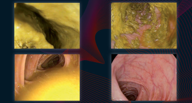
Typ 2 – Charakteristisch für Adenom
Farbe: dunkler (brauner) als Umgebung
Gefäße: zentral hellerer Bereich, umgeben von dickeren braunen Gefäßen


Oberfläche: oval, tubulär, gyrös — Vorhandensein von Tubuli, linear oder als Konvolut, zentral hellerer Bereich, umgeben von braunen Gefäßen.


Weitere Beispiele:


Typ 3 – Charakteristisch für Malignom (SM)
Farbe: dunkler als die Umgebung?, bräunlich, manchmal mit helleren Flecken
Gefäße: Areale von unterbrochenen oder fehlenden Gefäßen
Oberfläche: Amorphes oder kein Oberflächenmuster


NICE-Klassifikation
| Type 1 | Type 2 | Type 3 | |
|---|---|---|---|
| Color | Same or lighter than background | Brown relative to background (verify color arises from vessels) | Brown to dark brown relative to background; sometimes patchy whiter areas |
| Vessels | None, or isolated lacy vessels may be present coursing across the lesion | Brown vessels surrounding white structures* | Has area(s) of disrupted or missing vessels |
| Surface patterns | Dark or white spots of uniform size, or homogeneus absence of pattern | Oval, tubular or branched white structures* surrounded by brown vessels | Amorphous or absent surface pattern |
| Most likely pathology | Hyperplastic | Adenoma** | Deep submucosal invasive cancer |
| Sample Image |  |
 |
 |
| This classification can be applied using colonoscopes both with or without optical (zoom) magnification. * These structures (regular or irregular) may represent the pits and the epithelium of the crypt opening. ** Type 2 consists of Vienna classification types 3, 4 and superficial 5 (all adenomas with either low or high grade dysplasia, or with superficial submucosal carcinoma). The presence of high grade dysplasia or superficial submucosal carcinoma may be suggested by an irregular vessel or surface pattern, and is often associated with atypical morphology (e.g., depressed area). |
|||
Literaturverzeichnis
- Hewett DG, Kaltenbach T, Sano Y, Tanaka S, Saunders BP, Ponchon T, Soetikno R, Rex DK. Validation of a simple classification system for endoscopic diagnosis of small colorectal polyps using narrow-band imaging. Gastroenterology. 2012 Sep;143(3):599-607.e1. doi: 10.1053/j.gastro.2012.05.006. Epub 2012 May 15.
- Rex DK, Kahi CJ, Michael O’Brien M, et al.: The American Society for Gastrointestinal Endoscopy PIVI on real-time endoscopic assessment of the histology of diminutive colorectal polyps. Gastrointest Endosc 2011; 73:419-422
- McGill SK, Evangelou E, Ioannidis JP, Soetikno RM, Kaltenbach T. Narrow band imaging to differentiate neoplastic and non-neoplastic colorectal polyps in real time: a meta-analysis of diagnostic operating characteristics. Gut. 2013 Dec;62(12):1704-13. doi: 10.1136/gutjnl-2012-303965. Epub 2013 Jan 7
- Nagorni A, Bjelakovic G, Petrovic B. Narrow band imaging versus conventional white light colonoscopy for the detection of colorectal polyps. Cochrane Database Syst Rev. 2012 Jan 18;1:CD008361. doi: 10.1002/14651858.CD008361.pub2.
- van den Broek FJ, Reitsma JB, Curvers WL, Fockens P, Dekker E. Systematic review of narrow-band imaging for the detection and differentiation of neoplastic and nonneoplastic lesions in the colon (with videos). Gastrointest Endosc. 2009 Jan;69(1):124-35. doi: 10.1016/j.gie.2008.09.040.
- Wanders LK, East JE, Uitentuis SE, Leeflang MM, Dekker E. Diagnostic performance of narrowed spectrum endoscopy, autofluorescence imaging, and confocal laser endomicroscopy for optical diagnosis of colonic polyps: a meta-analysis. Lancet Oncol. 2013 Dec;14(13):1337-47. doi: 10.1016/S1470-2045(13)70509-6. Epub 2013 Nov 13
- Rastogi A, Bansal A, Wani S, et al. Narrow-band imaging colonoscopy–a pilot feasibility study for the detection of polyps and correlation of surface patterns with polyp histologic diagnosis. Gastrointest Endosc. 2008;67:280-6. Epub 2007 Dec 26.
- Rastogi A, Keighley J, Singh V, et al. High accuracy of narrow band imaging without magnification for the real-time characterization of polyp histology and its comparison with high-definition white light colonoscopy: a prospective study. Am J Gastroenterol. 2009;104:2422-30. Epub 2009 Jul 7.
- Rastogi A, Early DS, Gupta N et al.. Randomized, controlled trial of standard-definition white-light, high-definition white-light, and narrow-band imaging colonoscopy for the detection of colon polyps and prediction of polyp histology. Gastrointest Endosc 2011;74:593-602
- Gupta N, Bansal A, Rao D, et al. Accuracy of in vivo optical diagnosis of colon polyp histology by narrow-band imaging in predicting colonoscopy surveillance intervals. Gastrointest Endosc. 2012;75:494-502. Epub 2011 Oct 26
- Hassan C, Pickhardt PJ, Rex DK. A resect and discard strategy would improve cost-effectiveness of colorectal cancer screening. Clin Gastroenterol Hepatol. 2010;8:865-9, 869.e1-3. Epub 2010 Jun 1.
- Ignjatovic A, East JE, Suzuki N, er al. Optical diagnosis of small colorectal polyps at routine colonoscopy (Detect InSpect ChAracterise Resect and Discard; DISCARD trial): a prospective cohort study. Lancet Oncol. 2009;10:1171-8. Epub 2009 Nov
- Wallace MB, Kiesslich R. Advances in endoscopic imaging of colorectal neoplasia. Gastroenterology. 2010;138:2140-50.
- Ladabaum U, Fioritto A, Mitani A, et al.: Real-Time Optical Biopsy of Colon Polyps With Narrow Band Imaging in Community Practice Does Not Yet Meet Key Thresholds for Clinical Decisions. Gastroenterology. 2013 Jan;144(1):81-91. doi: 10.1053/j.gastro.2012.09.054. Epub 2012 Oct 3.
- Schachschal G, Mayr M, Treszl A, Balzer K, Wegscheider K, Aschenbeck J, Aminalai A, Drossel R, Schröder A, Scheel M, Bothe CH, Bruhn JP, Burmeister W, Stange G, Bähr C, Kießlich R, Rösch T. Endoscopic versus histological characterisation of polyps during screening colonoscopy.Gut. 2014 Mar;63(3):458-65. doi: 10.1136/gutjnl-2013-304562. Epub 2013 Jun 28.
Ähnliche Beiträge
BBPS
Verschiedene Maßnahmen haben die Mortalität des kolorektalen Karzinoms in den
KLASSIFIKATION ANSEHEN
Kontrollintervalle nach Polypektomie im Kolon
Nachsorgeintervalle nach Polypektomie Die Koloskopie ist das zuverlässigste Verfahren zur
KLASSIFIKATION ANSEHEN