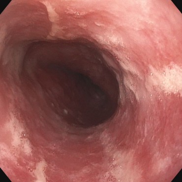
Image Challenge
 CIRS
CIRS
Question:
A colonoscopy, with a polypectomy in the ascending colon, was carried out in the hospital. The patient was also known to be suffering from chronic lymphatic leukemia, which was being treated with dasatinib. Following the intervention (approx. 3 hours later), a fall in hemoglobin from 11 g/dL to 9 g/dL was noted. At this point, the patient had a stable circulation and no pain; a digital rectal examination did not reveal any blood, and the abdomen was relaxed. The next check-up was carried out a further 3 hours later. On the way to the toilet, the patient had in the meantime passed into a state approaching collapse, with hypotension and tachycardia, and the Hb value had fallen to 8.0 g/dL. There was still no blood in the ampulla, but the abdomen was now tender on pressure.
The patient was transferred to the intensive-care unit for circulatory stabilization (volume substitution, catecholamines, administration of packed red blood cells). Colonoscopy was carried out. No relevant secondary bleeding was found in the colon, but a rigid abdomen was suddenly noted during splinting by the assistants. Ultrasound showed free fluid in the abdomen, and surgeons were immediately called. An emergency laparotomy was carried out, which revealed a splenic rupture.
What happend?
Feed back of our CIRS team:
False tract — splenic rupture after endoscopy
This is an interesting case that illustrates a kind of “blinker phenomenon” that may occur more often in shift situations.
When a hemoglobin drop with circulatory effects occurs after a polypectomy, secondary bleeding initially seems a more obvious explanation. In view of the amount of blood involved in this case, however, one would expect hematochezia to occur. In cases of unclear hemoglobin falls after colonoscopy, ultrasound should therefore always be used for orientation to look for free fluid and splenic injury. This is all the more important if, as in this case, the patient has already reported pain, since secondary bleeding does not usually cause any pain. Possibly making the situation worse is the fact that splenomegaly, potentially increasing the risk of splenic rupture, might have been present in this patient with known chronic lymphatic leukemia.
Splenic injury is a rare but typical complication of colonoscopy. According to the literature, the risk for splenic injury during the procedure is 1–21 cases per 100,000 colonoscopies per year [1, 2]. Marked tortuosity and loop formation in the bowel, with the resulting need for straightening and splinting maneuvers during the examination, as well as adhesions between the spleen and colon, have been reported to be risk factors for splenic injury [2–6]. However, splenic injury may also occur during uncomplicated examinations, only becoming evident days after the endoscopy.
Whether or not spleen-preserving management is possible must be checked in any case of splenic trauma. The current Level 3 guideline on “Multiple Trauma/Severe Injury — Treatment” recommends a conservative approach with intensive-care monitoring if the patient is hemodynamically stable and responds positively to volume substitution. Additional intra-abdominal injuries must be excluded using CT scanning [7].
During the informed consent discussion before the endoscopic procedure, the patient must be informed about the risk of injury to the spleen, and this point must be documented in writing. The patient must also be informed about what to do if symptoms develop (contacting or returning to the practice or hospital).
Modified American Association for the Surgery of Trauma (AAST) / Moore classification of splenic injury into five types:
| Type I | Subcapsular hematoma / capsular rupture | Usually conservative therapy |
| Type II | Type II Injury to the capsule and parenchyma, with no damage to segmental arteries | Usually spleen-preserving surgery / vascular intervention |
| Type III | Injury to the capsule, parenchyma, and segmental arteries | Usually spleen-preserving surgery / vascular intervention |
| Type IV | Injury to the capsule, parenchyma, and segmental or hilar vessels, detachment of the vascular pedicle | Usually splenectomy |
| Type V | Avulsion of the organ in the splenic hilum, with devascularization (interruption of the vascular supply) | Usually splenectomy |
References
- Kamath AS, Iqbal CW, Sarr MG, Cullinane DC, Zietlow SP, Farley DR, Sawyer MD. Colonoscopic splenic injuries: incidence and management. J Gastrointest Surg. 2009;13:2136–2140.
- Singla S, Keller D, Thirunavukarasu P, Tamandl D, Gupta S, Gaughan J, Dempsey D. Splenic injury during colonoscopy–a complication that warrants urgent attention. J Gastrointest Surg. 2012;16:1225–1234.
- Singla S, Keller D, Thirunavukarasu P, Tamandl D, Gupta S, Gaughan J, Dempsey D. Splenic injury during colonoscopy–a complication that warrants urgent attention. J Gastrointest Surg. 2012;16:1225–1234.
- Ha JF, Minchin D. Splenic injury in colonoscopy: a review. Int J Surg. 2009;7:424–427.
- Wherry DC, Zehner H. Colonoscopy-fiberoptic endoscopic approach to the colon and polypectomy. Med Ann Dist Columbia. 1974;43:189–192.
- Rex DK. Colonoscopic splenic injury warrants more attention. Gastrointest Endosc. 2013;77:941–943.
- Zandonà C, Turrina S, Pasin N, De Leo D. Medico-legal considerations in a case of splenic injury that occurred during colonoscopy. J Forensic Leg Med. 2012;19:229–233.
- S3 Leitlinie Polytrauma/ Schwerverletzten- Behandlung, AWMF Register-Nr. 012/019