
By loading the video, you accept YouTube's privacy policy.
Learn more
Sequenzen:

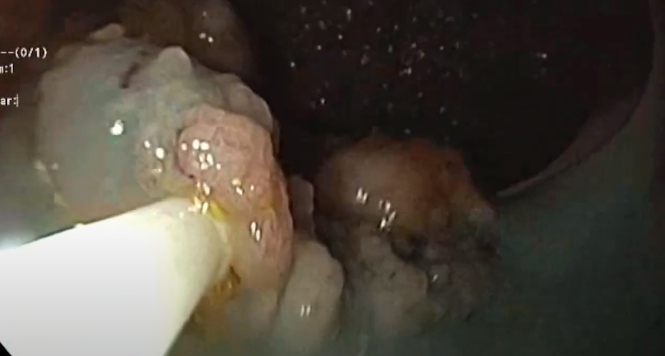
Diagnosis of a granular-type laterally spreading tumor at the right colic flexure.


Excision using the piecemeal technique with a 15-mm (monofilament) snare

Excision on the right edge of the tumor, where adjustment is difficult, and at the distal margin of the tumor. Positioning of the snare using the hypomochlion (fulcrum) technique.

Excision of residual tumor tissue after repeated injection underneath the lesion, and excision of residual pieces of the adenoma on the right margin of the tumor.

Demonstration of the resection margins and clip closure of the resection site with visible vessels.
Related Posts
Piecemeal EMR rectum
Piecemeal endoscopic mucosal resection (EMR) of an extensive laterally spreading
WATCH THE VIDEO
Sydney classification- assessment of deep mural injury after endoscopic mucosal resection.
Classification presented by Burgess NG et al. based on retrospective
VIEW CLASSIFICATION