Welcome to our image of the week!
Every week we present an endoscopic picture - click on the image for the diagnosis.
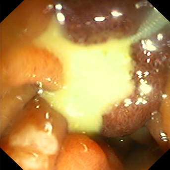
24. September 2019
diagnosis
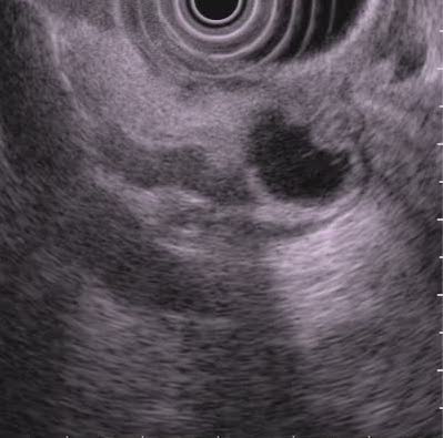
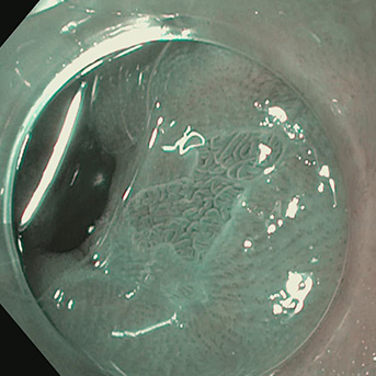
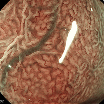
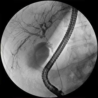
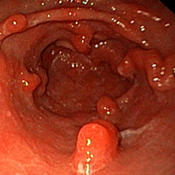
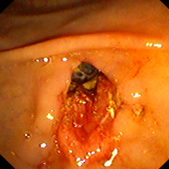
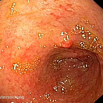
Endosonographic image of a duodenal diverticulum using watre filling
17. September 2019
diagnosis
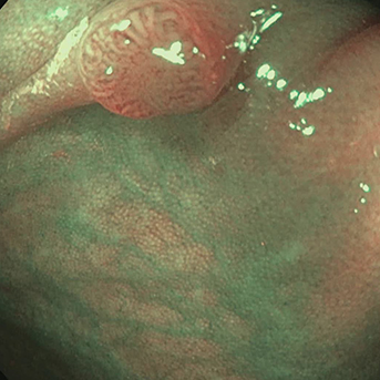
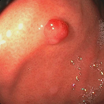
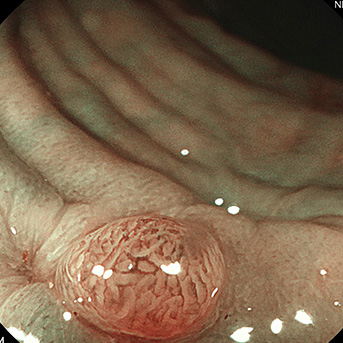
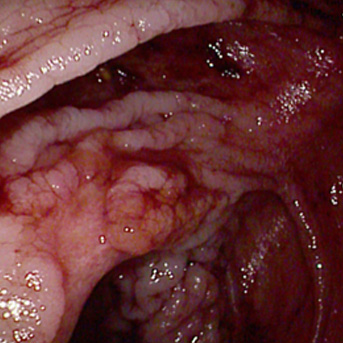
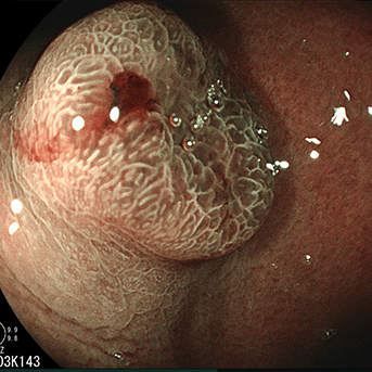
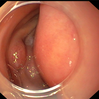
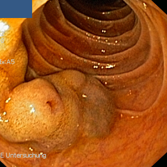
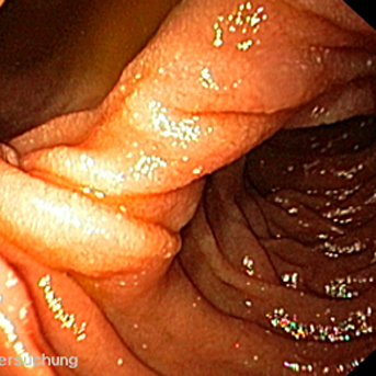
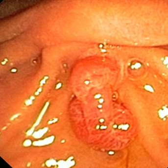
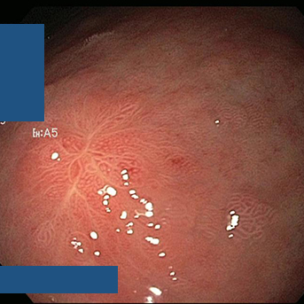
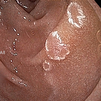
Small pedunculated sigmoid adenoma (low grade); the distorted pattern at the polyp tip does not indicate more severe histopathology
10. September 2019
diagnosis
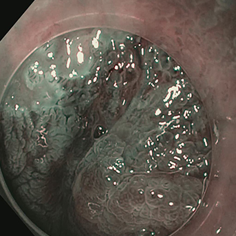
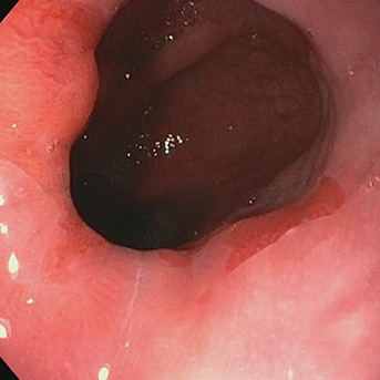
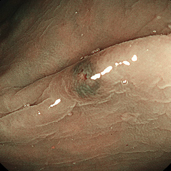
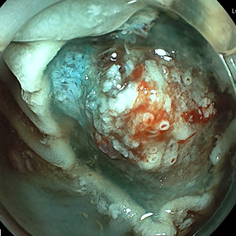
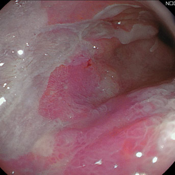
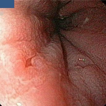
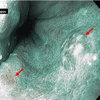
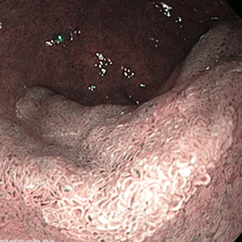
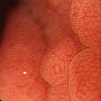
Close-up image of a Barrett carcinoma on NBI only visible with a cap
3. September 2019
diagnosis
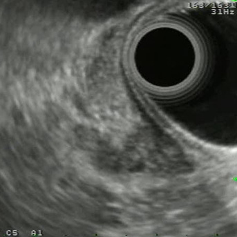
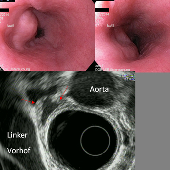
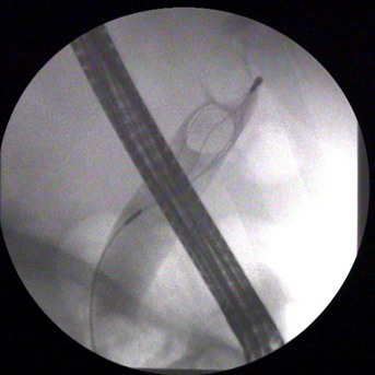
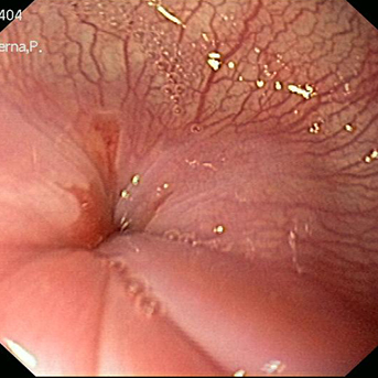
Endosonographic visualization of a distal bile duct carcinoma with echopoor and irregular wall thickening and a small part of the bile duct lumen
27. August 2019
diagnosis
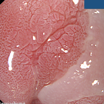
See image of last week – visualization with cap and acetic acid staining – typical Barrett epithelium
20. August 2019
diagnosis
Image of the cardia with questionable small Barrett island – see image next week
13. August 2019
diagnosis
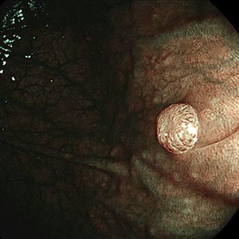
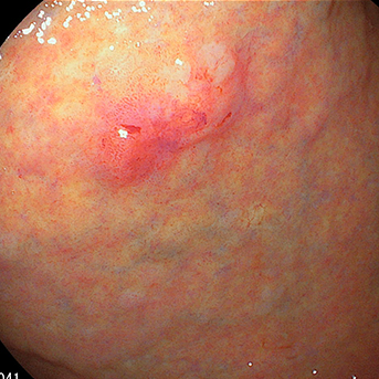
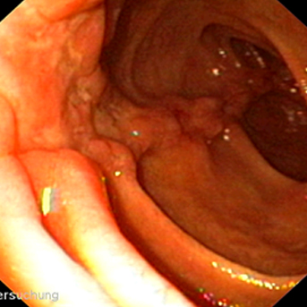
Reddish fundic gland polyp in the stomach (histologically confirmed)
6. August 2019

diagnosis
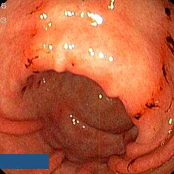
Slightly prominent appendiceal orifice in cecum
30. July 2019
diagnosis
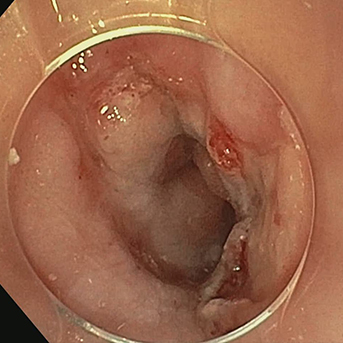
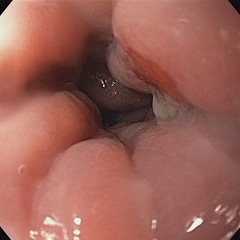
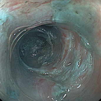
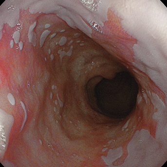
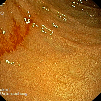
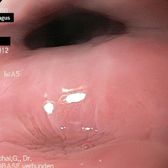
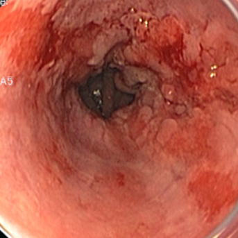
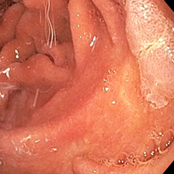
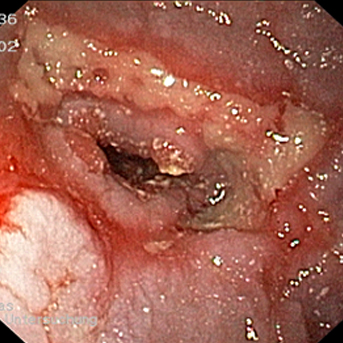
Distal esophagus in a patient with longstanding achalasia showing erosive changes, probably due to stasis
23. July 2019
diagnosis
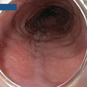
Valley Sign of a colon adenoma, first described by Dr. Rex in 2017
16. July 2019
diagnosis
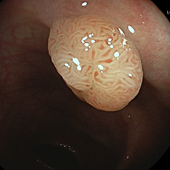
Tubullovillous adenoma.
9. July 2019
diagnosis
BLI highlights the mucosal surface and vascular pattern morphology. In combination with an optical zoom, in vivo diagnosis of a tubular adenoma can be easily achieved
25. June 2019
diagnosis
Submucosal tunnel during a POEM procedure.
18. June 2019
diagnosis
Post-inflammatory polyps in ulcerative colitis imaged with LCI. Identification of dysplasia in those cases can become extremely challenging.
11. June 2019
diagnosis
NET of the rectum imaged with BLI. Although small, the lesion can be clearly demarked for subsequent endoscopic resection.
28. May 2019
diagnosis
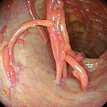
Multiple vessels after polypectomy. Not that endoscopic clipping is not always necessary.
21. May 2019

diagnosis
Mucomyotomy of Zenker’s diverticulum. Muscle fibers are already clearly exposed.
14. May 2019
diagnosis
Gastric cancer imaged with BLI for visualization of the mucosal surface and vascular pattern network. Endoscopic resection (ESD) was performed for the lesions.
7. May 2019
diagnosis
Long segment Barrett’s esophagus imaged with LCI.
30. April 2019
diagnosis
Small early gastric cancer visualized with LCI. The so called “red-in-purple” sign is always suspicious for neoplasia.
23. April 2019

diagnosis
Inflammatory colon polyps are common. As identification of dysplasia in those lesions is difficult, endoscopic resection should be performed.
16. April 2019
diagnosis
BLI highlights the mucosal surface and vascular pattern morphology. In combination with an optical zoom of 135-fold, optical diagnosis of a tubular adenoma can be easily achieved.
9. April 2019

diagnosis
Endoscopic full thickness resection with the FTRD system. Duplication of the single layers is clearly visible.
2. April 2019
diagnosis
Barrett’s cancer imaged with Linked Color Imaging (LCI) which strongly highlights the neoplastic tissue.
26. March 2019
diagnosis
Large duodenal lipoma
12. March 2019
diagnosis
Submucosal bulge in the esophagus, collapsing (right) suggesting esophageal duclication. Confirmation by endosonography showing mucosal structure in the wall of the duplication (below).
5. March 2019
diagnosis
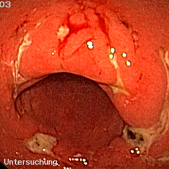
Duodenum in Graft versus Host Disease (GvHD)
19. February 2019
diagnosis
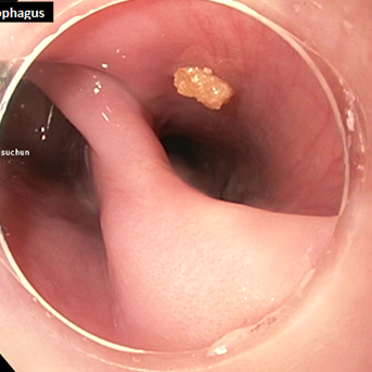
Food residues in a smaller Zenker diverticulum
12. February 2019
diagnosis
Duodenal varices
5. February 2019
diagnosis
Recurrent Zenker diverticulum, scarring and deviating
15. January 2019

diagnosis
Healed duodenal ulcer without fibrin
8. January 2019
diagnosis
Small Zenker diverticulum (1 cm on barium swallow), hardly visible on endoscopy beside a hypertrophic upper sphincter
25. December 2018

diagnosis
Healing duodenal ulcer with elevated margin at the upper duodenal curve
11. December 2018

diagnosis
Linitis plastica (diffusely infltrating gastric cancer)
4. December 2018
diagnosis
Duodenal bulb ulcer
27. November 2018
diagnosis
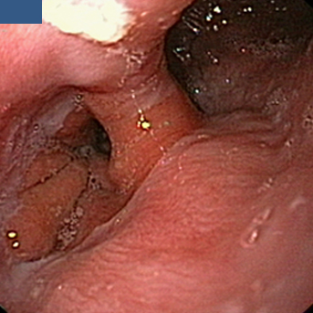
Epiphrenic diverticulum in the distal esophagus
20. November 2018
diagnosis
Early gastric cancer at the angular fold (incisura angularis)
13. November 2018
diagnosis
Patient with a known ulcerative colitis and elevated liver enzyms - ERCP with typical aspect of a PSC
6. November 2018
diagnosis
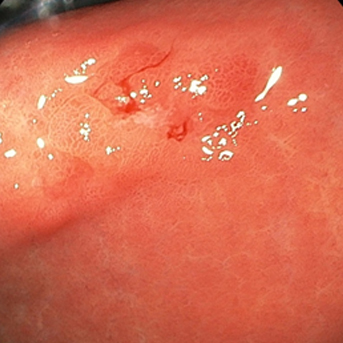
Barrett esophagus after forceps biopsies, simulating erosions or neoplasia
2. October 2018
diagnosis
Patient with a large common bile duct stone
25. September 2018
diagnosis
Duodenal diverticulum
18. September 2018
diagnosis
Flat erosive Barrett cancer (AEG cancer type I, AEG=adenocarcinoma of esophagogastric junction)
11. September 2018

diagnosis
Classic long-segment Barrett esophagus
4. September 2018
diagnosis
Capsule endoscopy in a patient with "Blue rubber bleb naevus Syndrom", showing a venous ectasia in the jejunum
28. August 2018

diagnosis
Two angiodysplastic lesions at the gastric incisura (angular fold)
21. August 2018
diagnosis
Patient with a pseudomembranous colitis (Clostridium difficile)
14. August 2018

diagnosis
Barrett esophagus with prominent vessels in a patient with portal hypertension
7. August 2018
diagnosis
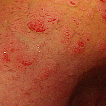
Multifocal areas of intestinal metaplasia (NBI imaging)
31. July 2018
diagnosis
Small flat duodenal adenoma in the descending duodenum
24. July 2018
diagnosis
Patient with Crohn's disease and fistulation from sigmoid colon to descending colon
17. July 2018
diagnosis
Known Barrett esophagus with 2 small neoplastic lesions, on histology metastases of breast cancer
10. July 2018
diagnosis
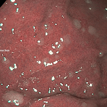
Fibrinous gastric erosions (NSAID)
3. July 2018
diagnosis
Flat duodenal adenoma at the distal end of the bulb
26. June 2018
diagnosis
Barrett mucosa in near focus and with acetic acid staining with central high-grade dysplastic lesion
19. June 2018
diagnosis
Pedunculated ampullary adenoma
5. June 2018
diagnosis
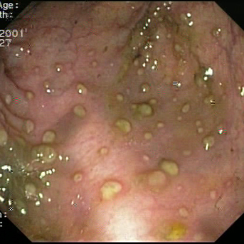
Polypoid gastric erosions
15. May 2018
diagnosis
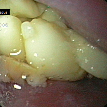
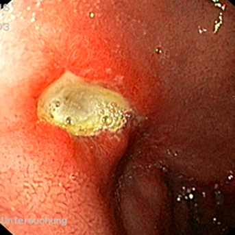
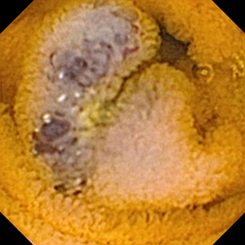
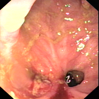
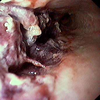
Esophagus in advanced achalasia showing chronically inflamed and thickened mucosa and a distal ulcer due to pressure necrosis by obstructing food
1. May 2018
diagnosis
Hämorrhagic gastric erosions
24. April 2018
diagnosis
Duodenal involvement in Crohn´s disease
10. April 2018
diagnosis
Esophagus in achalasia showing muscular rings and prominent veins
3. April 2018
diagnosis
Perforation after endoscopic sphincterotomy
27. March 2018
diagnosis
Gastric erosion with biopsy positive for cancer, but ESD specimen negative („biopsy cancer“)
20. March 2018
diagnosis
Duodenal bulb with gastric metaplasia (magnification endoscopy)
6. March 2018
diagnosis
Distal esophagus in achalasia; muscular spasm leads to overvisualization of submucosal vessels
27. February 2018
diagnosis
Swollen und reddened papille with massive pus evaculation
20. February 2018
diagnosis
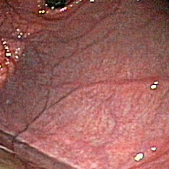
Atrophic gastritis in the gastric fundus with vascular pattern
13. February 2018
diagnosis
Duodenal bulb with reddisch gastric metaplasia
6. February 2018
diagnosis
Boerhave syndrome with deep defect.
30. January 2018

diagnosis
Papilla in a patient with Billroth II anatomy and pus
23. January 2018
diagnosis
Atrophic gastritis with small neuroendocrine tumor
15. January 2018
diagnosis
Duodenal mucosa with 3 small metastases of breats cancer (histologically confirmed)
